Ultrasonic blood vessel measurement apparatus and method

a measurement apparatus and ultrasonic technology, applied in the field of ultrasonic blood vessel measurement apparatus and method, can solve the problems of arterial wall rupture, heart attack, stroke, embolism, etc., and achieve the effect of accurately representing the lumen/intima boundary, reducing error, and reducing the risk of strok

Active Publication Date: 2010-06-01
FUJIFILM SONOSITE
View PDF63 Cites 37 Cited by
  • Summary
  • Abstract
  • Description
  • Claims
  • Application Information

AI Technical Summary

Benefits of technology

[0014]It is another object of embodiments of the present invention to reduce error in measurements by restricting searches for the tissue boundaries, such as the lumen / intima boundary and media / adventitia boundary, to regions likely to contain them.
[0043]In another embodiment, once a tissue structure aspect density is determined, the density values can be normalized to remove any dependency on the instrument gain setting when the imaging device, e.g., ultrasound device, captured the tissue structure image. This can be accomplished by normalizing the density values obtained by both 1) The minimum average intensity of the image (which typically corresponds to the lumen of the carotid artery when the image is of the carotid artery), and / or 2) The maximum average intensity of the image (which typically corresponds to the adventitia of the far wall of the carotid artery when the image is of the carotid artery).

Problems solved by technology

CAD may cause shortness of breath, angina, or even heart attack.
The buildup of plaque may also create a risk of stroke, heart attacks, and embolisms caused by fragments of plaque detaching from the artery wall and occluding smaller blood vessels.
The risk of arterial wall rupture and detachment of portions of the blood clot covering the rupture is particularly great shortly after blood clot formation when it is soft and easily fragmented at that stage.
Measurement of the atherosclerotic burden of the coronary artery itself is difficult and invasive.
This assessment therefore involves multiple invasive procedures over time.
This method of forming images is subject to limitations and errors.
For example, images may be subject to noise from imperfect sensors.
Another source of error is the attenuation of sound waves that reflect off tissue located deep within the body or beneath denser tissue.
Random reflections from various objects or tissue boundaries, particularly due to the non-planar ultrasonic wave, may add noise also.
The limitations of ultrasonography complicate the interpretation of ultrasound images.
Such systems may reject an entire column of image data in which selected portions of the wall are not readily identifiable.
This method fails to take advantage of other portions of the artery wall that are recognizable in the column.
Furthermore, examining columns of pixels singly fails to take advantage of accurate information in neighboring columns from which one may extrapolate, interpolate, or otherwise guide searches for information within a column of pixels.
Another limitation of prior methods is that they fail to adequately limit the range of pixels searched in a column of pixels.
Noise and poor image quality can cause any search for maxima, minima, or intensity gradients to yield results that are clearly erroneous.

Method used


Image

  • Ultrasonic blood vessel measurement apparatus and method
  • Ultrasonic blood vessel measurement apparatus and method
  • Ultrasonic blood vessel measurement apparatus and method

Examples


Embodiment Construction

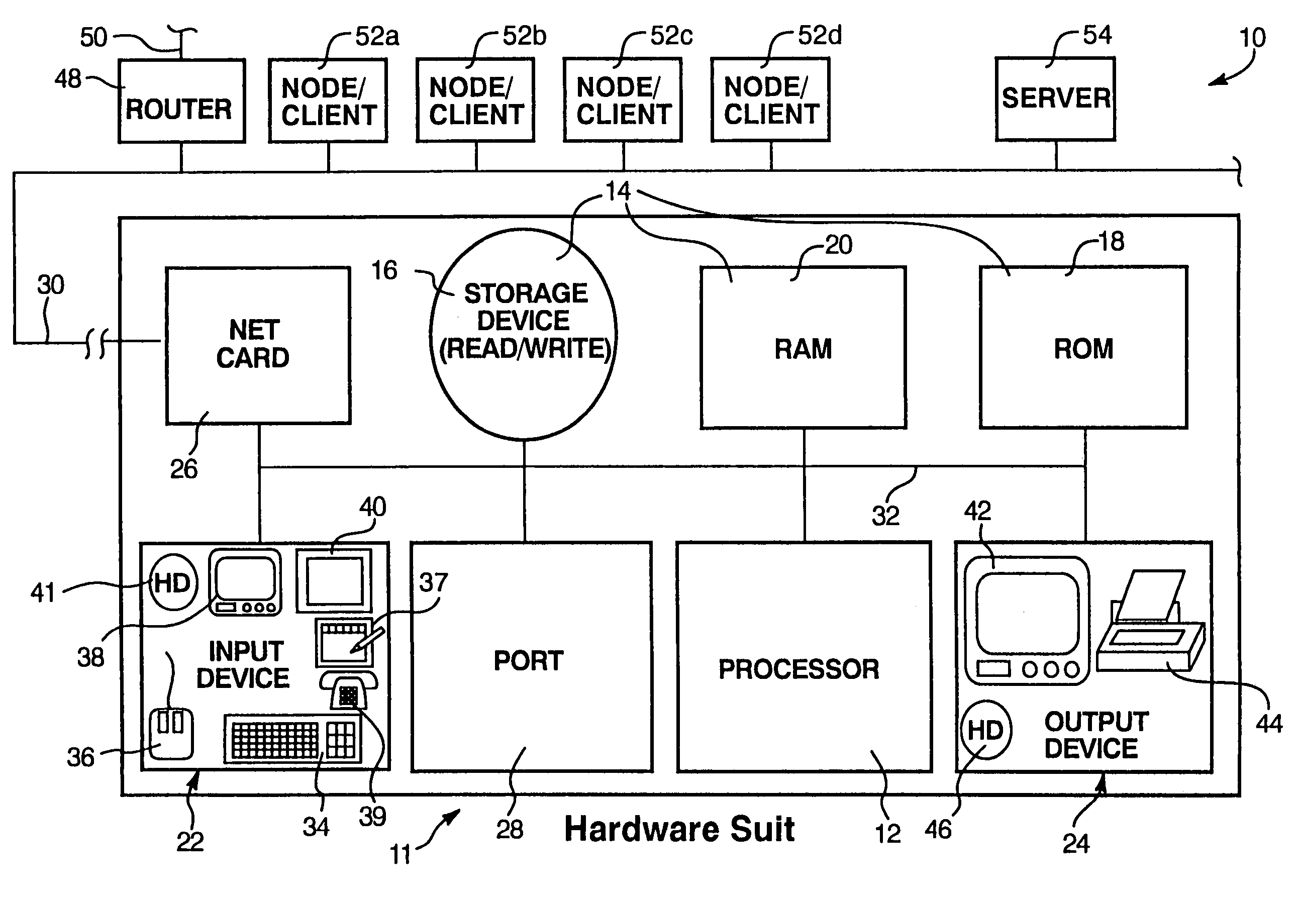
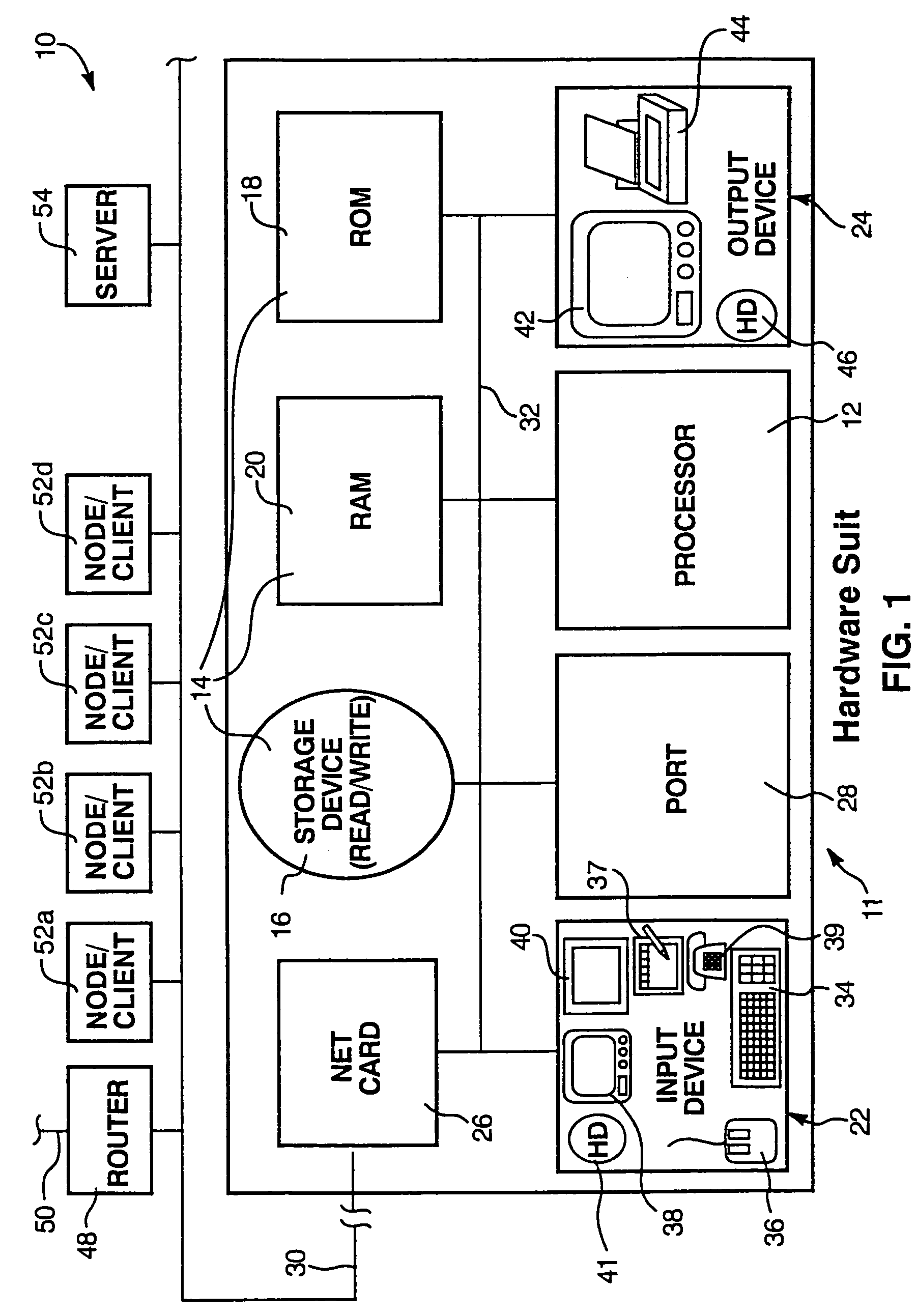
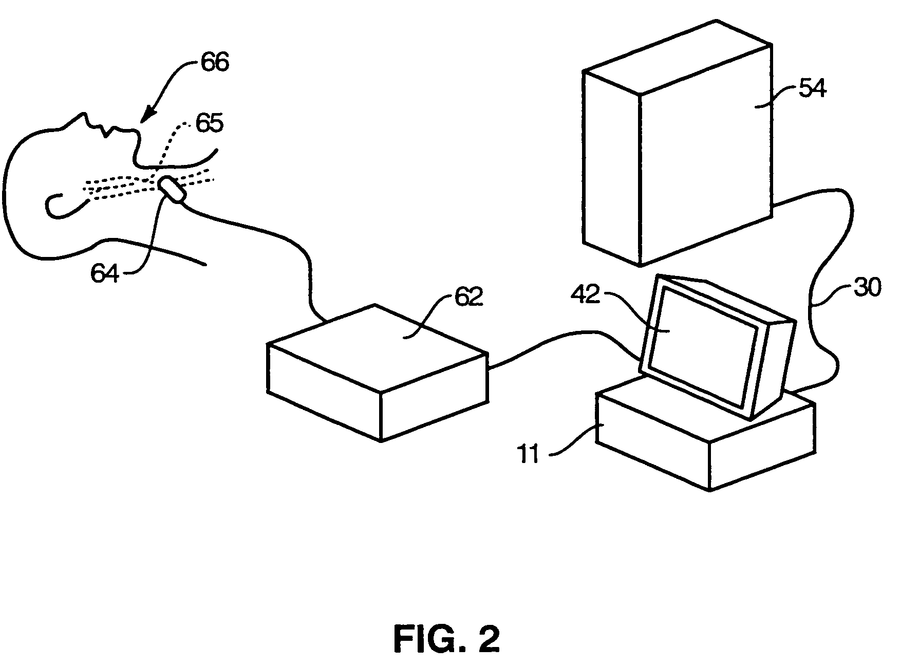
[0082]It will be readily understood that the components of the present invention, as generally, described and illustrated in the Figures herein, could be arranged and designed in a wide variety of different configurations. Thus, the following more detailed description of the embodiments of the system and method of the present invention, as represented in FIGS. 1-29, is not intended to limit the scope of the invention, as claimed, but is merely representative of certain presently preferred embodiments in accordance with the invention. These embodiments will be best understood by reference to the drawings, wherein like parts are designated by like numerals throughout.

[0083]Those of ordinary skill in the art will, of course, appreciate that various modifications to the details illustrated in FIGS. 1-29 may easily be made without departing from the essential characteristics of the invention. Thus, the following description is intended only by way of example, and simply illustrates certa...


PUM


Abstract

Disclosed are systems and methods for identifying various tissue structure aspects, such as boundaries of arterial walls, within an image, such as an ultrasound image. Various measurements may be made using information with respect to the identified aspects of tissue structure. For example, intima-media thickness measurements may be made. Additionally or alternatively, tissue structure aspects, such as plaque within an artery, may be characterized, such as by determining a density thereof. Various information, such as measurement datums, used in identification of aspects of tissue structure in one image may be stored and applied to subsequent images, such as images of a video sequence.

Description

CROSS-REFERENCE TO RELATED APPLICATIONS[0001]This application is a continuation-in-part of U.S. patent application Ser. No. 10 / 682,699 filed Oct. 9, 2003 entitled “ULTRASONIC BLOOD VESSEL MEASUREMENT APPARATUS AND METHOD,” which is a continuation-in-part of U.S. patent application Ser. No. 10 / 407,682 filed Apr. 7, 2002 entitled “METHOD, APPARATUS, AND PRODUCT FOR ACCURATELY DETERMINING THE INTIMA-MEDIA THICKNESS OF A BLOOD VESSEL;” and claims priority through the foregoing patent applications to U.S. provisional patent applications Ser. No. 60 / 424,027 filed Nov. 6, 2002 entitled “METHOD AND APPARATUS FOR INTIMA-MEDIA THICKNESS MEASURING MECHANISM EMBEDDED IN ULTRASOUND IMAGING DEVICE;” Ser. No. 60 / 424,464 filed Nov. 8, 2002 entitled “METHOD AND APPARATUS FOR MEASURING INTIMA-MEDIA THICKNESS ACROSS MULTIPLE SIMILAR IMAGES;” Ser. No. 60 / 424,471 filed Nov. 8, 2002 entitled “METHOD AND APPARATUS FOR INCORPORATING INTIMA-MEDIA TAPERING EFFECTS ON INTIMA-MEDIA THICKNESS CALCULATIONS;” Ser...

Claims


Application Information

Patent Timeline
Patent Type & Authority Patents(United States)
IPC IPC(8): A61B5/06A61B8/08G06T7/00G06T7/60
CPCA61B5/02007A61B5/1075A61B8/0858A61B8/463G06T7/0012G06T7/0083G06T7/606G06T2207/30101G06T2207/10132A61B8/565A61B8/0891G06T7/12G06T7/66
Inventor FRITZ, HELMUTHFRITZ, TERRY
Owner FUJIFILM SONOSITE
Why Eureka
Try Eureka