Method, system and storage medium which includes instructions for analyzing anatomical structures

a technology of anatomical structure and instructions, applied in the field of method, system and storage medium, can solve the problems of mci-related hippocampal hypometabolism invisible to the human observer, mci-related hippocampal metabolic abnormalities, and the use of cortical hypometabolism in identifying patients with mci is controversial, and achieves good comparability, high intra-rater reliability, and the effect of determining the anatomical validity

Inactive Publication Date: 2010-08-31
NEW YORK UNIV
View PDF14 Cites 19 Cited by
  • Summary
  • Abstract
  • Description
  • Claims
  • Application Information

AI Technical Summary

Benefits of technology

[0029]In particular. a first embodiment of the method, system and storage medium of the present invention is provided which allows for accurate sampling of the hippocampus on PET using automated (or computerized) routines. This was done by assessing the extent of hippocampus volume overlap after spatial normalization of elderly brains, as done with VBA techniques. Based on high resolution MR and large normative sample, the embodiment generally establishes a probabilistic map of the hippocampus in the stereotactic space. The map may be translated to a probabilistic masking image that may be used in a reliable and automated procedure for sampling the hippocampus on a PET scan. This method was then tested for reliability against the ROIs and for accuracy in diagnosing MCI and AD. This may demonstrate that when anatomically correct sampling is utilized the voxel-based method is reliable enough for the hippocampus.
[0031]Visual rating of the MTL on MRI scans have shown a good comparability with MRI volume study. Accordingly, visual rating of FDG-PET scans may permit (a) developing a reliable and anatomically valid visual rating scale of MTL hypometabolism on FDG-PET, (b) determining the anatomical validity of the visual MTL rating by comparing the diagnostic capacity of the MTL rating with quantitative metabolic values derived from the gold-standard ROI sampling technique, and (c) comparing the diagnostic accuracy of the MTL rating to the cortical rating in terms of being able to separate clinical groups.
[0032]Generally, the second exemplary embodiment of the method, system and storage medium of the present invention is capable of providing a high intra and inter-rater reliability for both the cortical and MTL ratings. Across several comparisons among raters, Intra-Class Correlation Coefficients (ICCs) ranged from 0.90 to 0.96 with p<0.001. Conventional cortical rating generally significantly discriminates AD from NL (in some cases, achieving 100% accuracy) and AD from MCI (in some cases, achieving 88% accuracy) but fails to distinguish MCI from NL. This embodiment may distinguish MCI from NL via the MTL rating (achieving, for example, a 74% accuracy), which may improve the early diagnosis of AD at the MCI stage over traditional methods. The MTL ratings may yield a diagnostic accuracy equivalent to so-called “gold-standard” ROI MRglc measures.

Problems solved by technology

The ROI method requires MR scans to guide the PET sampling and time-consuming manual outlining of the hippocampus.
Unfortunately, current implementations typically preserve only the maximum metabolic activity along each projection ray, thus rendering hippocampal hypometabolism invisible to the human observer.
However, few (if any) PET studies using voxel-based or surface projection methods report hippocampal metabolic abnormalities in MCI or AD as compared to controls.
As with the prior discussion of PET studies, such studies rely on estimation of changes in cortical brain metabolism and have not reported data on the hippocampus.
However, the usefulness of cortical hypometabolism in identifying patients with MCI is controversial.
Similarly, current methods and apparatuses for anatomically validating MTL hypometabolism may be unreliable or imprecise, especially when a visual evaluation is employed.
Computerized ROI implementations, however, may be both time-consuming and relatively expensive when compared to visual evaluations, and thus may be less suitable for diagnostic purposes.
However, these studies compared AD patients to normal controls and did not evaluate the accuracy of PET ratings in the diagnosis of MCI or to normal subjects who may be at increased future risk for MCI or AD.
However, like PET scans, SPECT scans have shown only a limited ability to examine a patient's hippocampus.

Method used


Image

  • Method, system and storage medium which includes instructions for analyzing anatomical structures
  • Method, system and storage medium which includes instructions for analyzing anatomical structures
  • Method, system and storage medium which includes instructions for analyzing anatomical structures

Examples


first exemplary embodiment

1. First Exemplary Embodiment

Hippocampal Mask (HipMask)

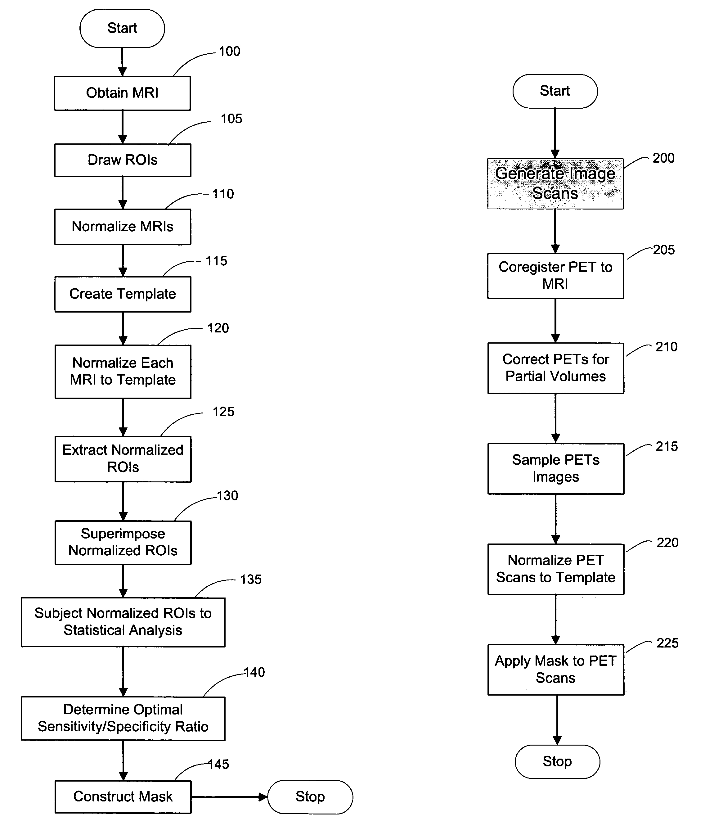
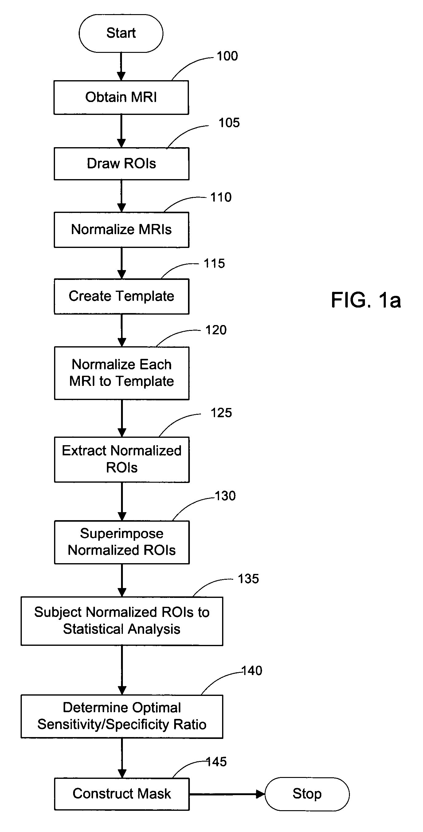
[0053]FIG. 1a depicts a flow diagram of an exemplary embodiment of a method for constructing a HipMask. FIG. 9 depicts an exemplary display of such exemplary method, in which images for the scanned cross-sectional view of a brain is displayed as MRI 10, Hippocampal ROI 20, and HipMask 30. Indeed, the HipMask 30 image can be constructed by utilizing the exemplary method illustrated in the flowchart of FIG. 1. Referencing the exemplary method shown in FIG. 1, after a group of patients of sufficient size are assembled (as discussed below), magnetic resonance images (MRIs) are taken of each patient's brain in operation 100 to create a set of MR scans or MRIs. (The terms “MR scan” and “MRI” are used interchangeably herein.) The MR scanning procedure may employ any scanning parameters that provide a sufficiently precise brain image. Briefly, one embodiment of the present invention may employ MR scans 10 generated by a-1.5 T General El...

second exemplary embodiment

5. Second Exemplary Embodiment

Visual Rating of Medial Temporal Lobes

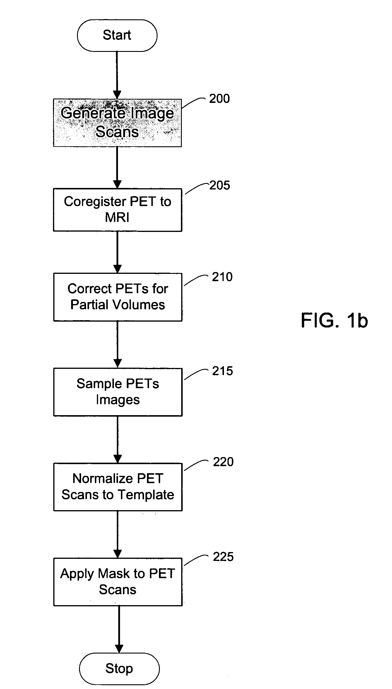
[0132]A second exemplary embodiment of the method, system and storage medium according to the present invention is provide for evaluating and analyzing brain tissue shown on the PET scans 60. Similar to the first exemplary embodiment, the second exemplary embodiment may be broadly applicable to evaluation and analysis of imaged brain tissue, such as that shown in the PET scan 60.

[0133]For example, according to this embodiment, a standardized MRI scan protocol can be conducted on a set of patients to provide a set of MRIs 10. The MRI scanning procedures have been previously described above, as well as in certain references known to those of ordinary skill in the art.

[0134]For example, the MRI scans may be acquired on a 1.5 T General Electric Signa imager (General Electric, Milwaukee, USA) using a T1-weighted fast-gradient-echo sequence with repetition time=35 ms, echo time=9 ms, and flip angle 60°. Images may be reco...


PUM


Abstract

A method, system and storage arrangement are provided for effectuating an evaluation and analysis of anatomical structures, and creating and / or modifying images associated therewith. In particular, at least two images associated with the anatomical structure can be normalized so as to produce normalized image. A normalized set of regions of interest can be obtained based on the normalized images. Each of the normalized set of regions of interest may be analyzed to provide analysis data. Further, the anatomical structure mask may be created and / or modified based on the analysis data. Another embodiment provides for rating and analyzing anatomical structures.

Description

CROSS-REFERENCE TO RELATED APPLICATION(S)[0001]The present application claims priority from U.S. Patent Application Ser. No. 60 / 588,4677 filed Jul. 16, 2004 and U.S. Patent Application Ser. No. 60 / 691,715 filed Jun. 16, 2005, the entire disclosures of which are incorporate herein by reference.FIELD OF THE INVENTION[0002]The present invention relates to a method, system and storage medium which includes instructions for analyzing anatomical structures (brain tissue and metabolism). More particularly, this analysis can include sampling and analysis of the hippocampus and medial temporal lobe, as well as an application of the sampling and analysis to assessing the presence of mild cognitive impairment and Alzheimer's disease, including in persons not yet displaying symptoms.BACKGROUND INFORMATION[0003]The following abbreviations will be used throughout the specification as follows:[0004]“AD”—Alzheimer's disease[0005]“MCI”—mild cognitive impairment[0006]“GDS”—global deterioration scale[...

Claims


Application Information

Patent Timeline
Patent Type & Authority Patents(United States)
IPC IPC(8): G06K9/00
CPCG06T7/0012G06T7/0022G06T7/0083G06T7/0087G06T2207/10088G06T2207/10104G06T2207/20104G06T2207/30016G06T7/97G06T7/12G06T7/143
Inventor DE LEON, MONY J.MOSCONI, LISATSUI, WAI-HONRUSINEK, HENRY
Owner NEW YORK UNIV
Why Eureka
Try Eureka