Echogenic needle catheter configured to produce an improved ultrasound image

a technology of ultrasound image and needle catheter, which is applied in the field of medical devices, can solve the problems of affecting the performance characteristics, strength, flexibility and softness of the distal tip, and metal additives can impair the ability to ultrasonically image the distal tip, so as to reduce the amount of metal in the tip, reduce or eliminate the effect of pyramid artifacts

Inactive Publication Date: 2010-09-14
ABBOTT CARDIOVASCULAR
View PDF77 Cites 11 Cited by
  • Summary
  • Abstract
  • Description
  • Claims
  • Application Information

AI Technical Summary

Benefits of technology

[0009]The ICPs readily blend or otherwise combine with the matrix polymer materials, and preferably possess relatively high thermal oxidative stability (e.g., up to about 300° C.) which facilitates processing the ICPs into the matrix polymer material without degradation or loss of conductivity. Consequently, the ICPs can be melt-processed with a variety of commonly used low-conductive matrix polymers, for ease of manufacturability and without the compounding difficulties of metal or carbon additives. Compared to conductive metal-filled systems, the ICPs provide substantial weight savings, flexibility, durability, low-temperature processibility, and tailored reproducible conductivity. Additionally, due to the improved compatibility with the matrix polymer, the ICPs preferably have reduced sloughing compared to carbon loaded formulations, and preferably do not disadvantageously affect the cohesive strength of the matrix polymer.
[0010]The ICP distal tip electrode facilitates ultrasonic imaging of the catheter distal end within the patient, compared to a conventional metallic tip electrode, by reducing the amount of metal in the distal tip. Specifically, metallic material in a conventional catheter distal tip absorbs stores and then reemits the sonic energy of the ultrasonic imaging device, causing the metal in the tip to ring like a bell, sending out ultrasonic energy until the sonic energy that it has stored is depleted. This absorbed, stored and then reemitted sonic energy is received by the ultrasonic imaging device and creates images behind the catheter tip that decrease in brightness and size as the stored sonic energy is depleted, forming the tip pyramid artifact. On the other hand, polymeric materials produce echoes from their surfaces in the body that are usually of less amplitude than the thick metallic surfaces of conventional electrode tips. Additionally, polymeric materials are generally more dissipative of sonic energy than metallic materials and thus, if any pyramid artifact is produced, it is of smaller amplitude than those produced by tips / electrodes formed completely of metal or of a higher proportion of metallic materials. A disadvantageously bright / long duration / large tip pyramid artifact obscures the actual image of the catheter tip and surrounding anatomy. In contrast, a distal tip of the invention, configured to minimize the amount of metallic material at the distal tip, reduces the amount of sonic energy that the tip stores and then reemits to thereby reduce the brightness and duration of the tip pyramid artifact. The soft polymeric distal tip completely eliminates, or at least reduces, the tip pyramid artifact of prior metallic or metallic / polymer hybrid electrode catheter tips.
[0012]As discussed above, the polymeric distal tip of the invention prevents or reduces the tip pyramid artifact image. However, in the absence of a direct echo from the tip, the only ultrasonic image of the tip may be that due to the absorbed, stored and then reemitted sonic energy (i.e., the tip pyramid artifact). As a result, in a presently preferred embodiment, the polymeric distal tip has a spherical shape, as set forth in copending, commonly assigned U.S. patent application Ser. No. 11 / 293,420 ('420 Application”), incorporated by reference herein in its entirety. The spherical distal tip shape is configured to produce direct echoes, from a large angular range, and thereby prevents or minimizes the potential for misreading the position of the distal tip from the ultrasonic image, by avoiding the absence of an imaged direct echo from the distal tip. As discussed in the '420 Application, the spherical distal tip shape produces diffuse echoes such that the distal tip is directly imaged over a range of angles (relative to the catheter) substantially greater than 180°. Thus, the conductive polymeric distal tip of the invention, provided with a spherical shape, facilitates accurately positioning the catheter distal tip under ultrasonic imaging by providing an imaged direct echo with little or no tip pyramid artifact over a wide range of angles (i.e., with the catheter tip oriented in the patient at a wide variety of angles relative to the ultrasonic imaging device outside of the patient). The imaged direct echo indicates the actual location of the distal tip in the patient, unlike a tip pyramid artifact (if present) which is located behind the actual location of the catheter tip due to the delay in reemitting the sonic energy in the direction of the imaging device.
[0015]In one embodiment, a method of the invention generally comprises advancing within a patient's anatomy an echogenic needle catheter comprising an elongated shaft, a polymeric distal tip which is formed at least in part of an ICP and which has a port at a distal end of the distal tip, and preferably a needle which extends distally from the distal tip port in an extended configuration. The method includes directing sonic energy at the distal tip from an ultrasonic imaging device, to produce an ultrasonic image of the distal tip with eliminated or reduced pyramid artifact compared to a distal tip having a metallic tip electrode, and positioning the distal tip at a desired location within the patient.
[0016]A catheter distal tip of the invention minimizes the amount of metal in the tip and thus reduces its echo amplitudes, and reduces or eliminates its pyramid artifact. The ICP conductive materials have improved electrical, mechanical and melt-flow properties. Specifically, a catheter distal tip of the invention formed of the ICP has an improved combination of good cohesive strength, flexibility, and high conductivity, unlike prior “conductive polymeric” distal tips which required high levels of metallic or carbon doping to be conductive. Additionally, the ICP conductive blends have greatly reduced compounding difficulties compared with the prior metal- or carbon-doped formulations. These and other advantages of the invention will become more apparent from the following detailed description and exemplary drawings.

Problems solved by technology

As a result, because metals and carbon are not polymers, they can disadvantageously affect the performance characteristics, such as the strength, flexibility and softness, of the distal tip.
Additionally, metal additives can impair the ability to ultrasonically image the distal tip as discussed in more detail below.

Method used


Image

  • Echogenic needle catheter configured to produce an improved ultrasound image
  • Echogenic needle catheter configured to produce an improved ultrasound image
  • Echogenic needle catheter configured to produce an improved ultrasound image

Examples


Embodiment Construction

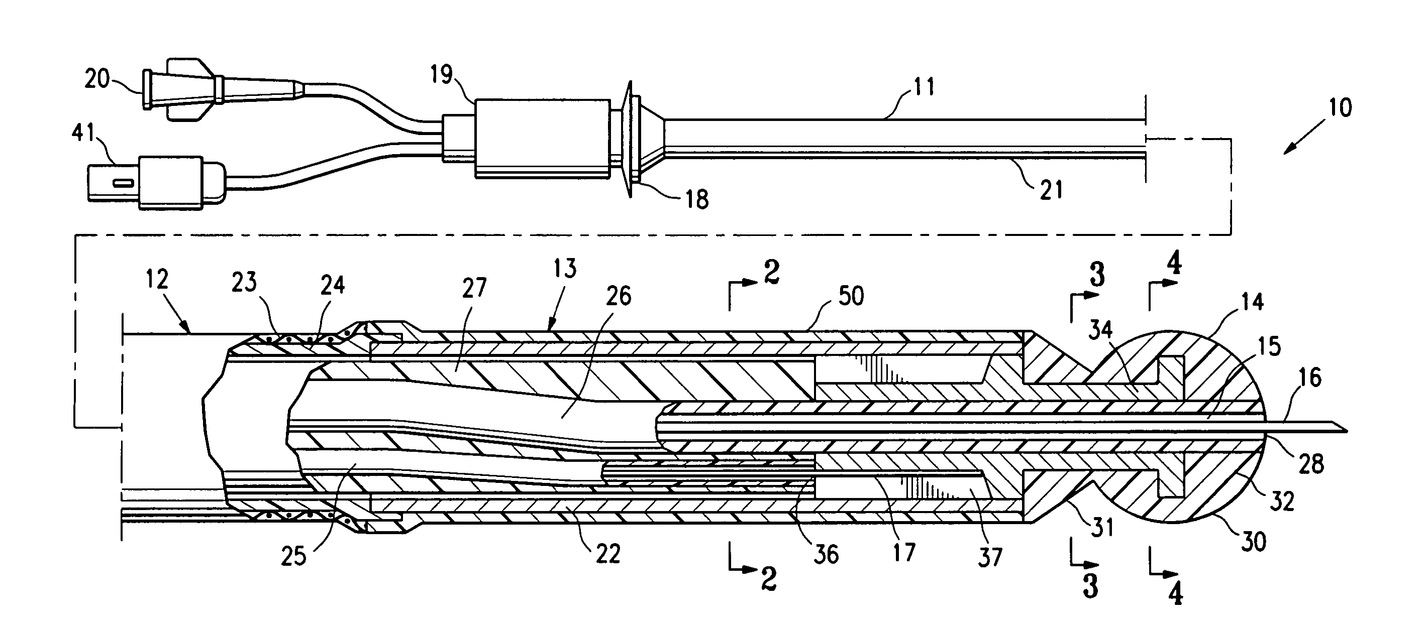
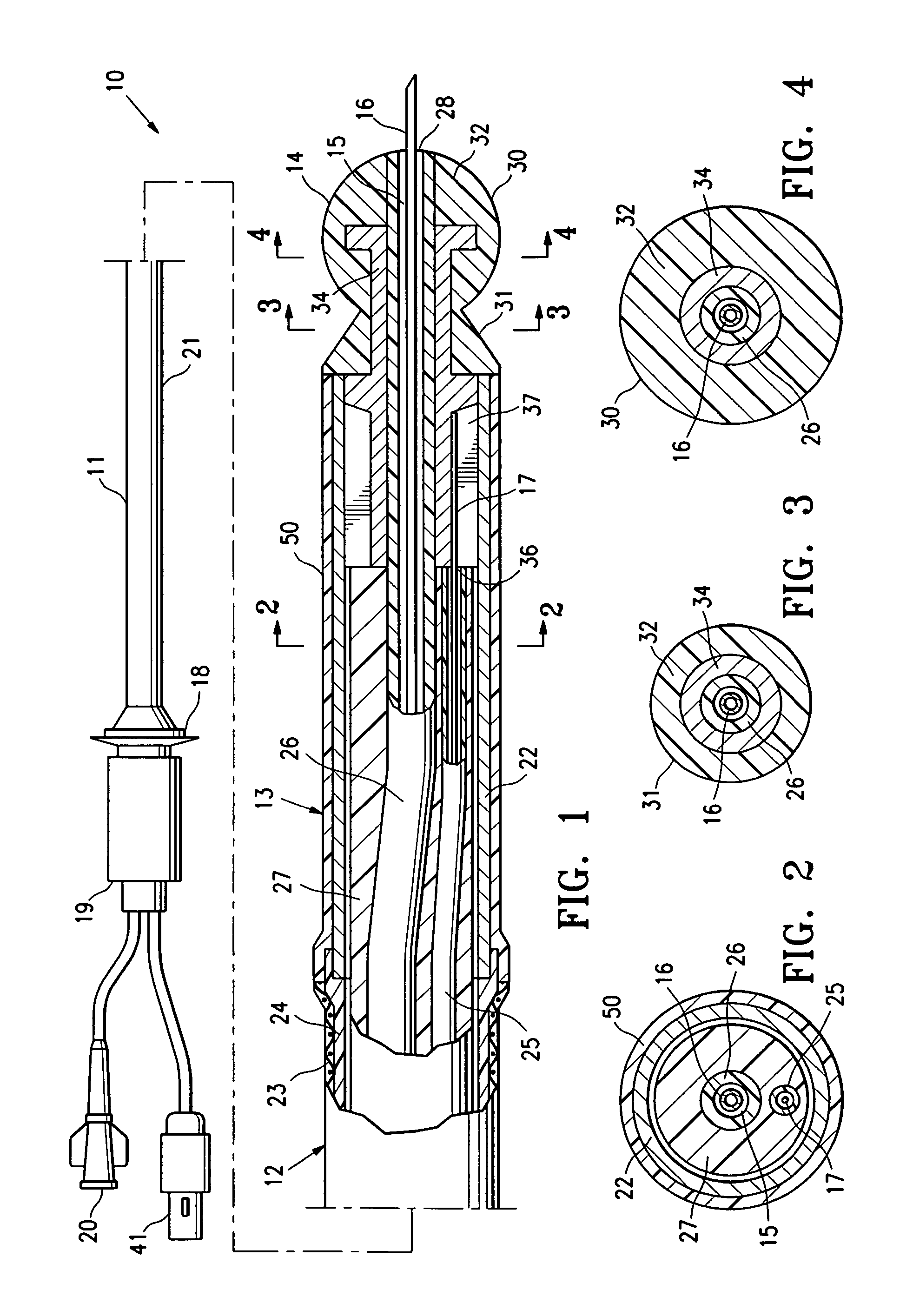
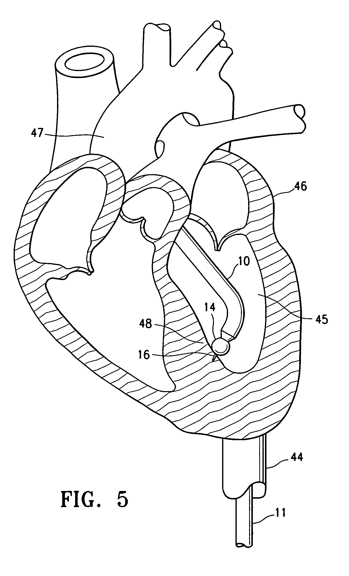
[0020]FIG. 1 illustrates a needle catheter 10 which embodies features of the invention. In the embodiment illustrated in FIG. 1, the needle catheter 10 generally comprises an elongated shaft 11 having a proximal shaft section 12, a distal shaft section 13, and a needle containing lumen 15, and a distal tip 14 at the distal end of the shaft 11. A needle 16 is slidably disposed within the lumen 15 of the shaft, with an extended configuration in which the needle distal end extends distally from the distal end of the catheter (see FIG. 1), and with a retracted configuration (not shown) in which the needle distal end is proximally retracted into the catheter lumen 15. In the illustrated embodiment, the catheter 10 has an elongated deflection member 17 (e.g., a tendon wire) connected to a deflection control mechanism 18 at a proximal adapter 19, for deflecting the distal end of the catheter 10. To effectively deflect the distal end of the catheter the deflection member 17 is preferably ne...


PUM

PropertyMeasurementUnit
circumference angleaaaaaaaaaa
conductivityaaaaaaaaaa

Abstract

An echogenic catheter, such as a needle catheter, formed at least in part of an intrinsically conductive organic polymer for providing a highly conductive surface in combination with an improved ability to ultrasonically image the catheter, and a method of performing a medical procedure using a catheter of the invention.

Description

BACKGROUND OF THE INVENTION[0001]The invention relates to the field of medical devices, and more particularly to echogenic catheters, such as needle catheters.[0002]An essential step in treating or diagnosing cardiac tissue or cardiovascular diseases using an interventional catheter is the proper placement of the catheter at a desired location within the patient, which consequently requires accurate imaging of the catheter location within the patient. Although various methods of imaging catheters within a patient are possible, ultrasonic imaging (also referred to as acoustic imaging) would provide several advantages. For example, ultrasonic imaging is very safe for the expected extended time periods required for therapy guidance, unlike CT / EBCT (Electron Beam Computed Tomography) or bi-planar fluoroscopy. Additionally, ultrasound is relatively inexpensive compared to other imaging modalities such as MRI or CT / EBCT, and can provide tissue diagnostics such as wall motion and thickness...

Claims


Application Information

Patent Timeline
Patent Type & Authority Patents(United States)
IPC IPC(8): A61B8/14
CPCA61B5/0402A61B8/06A61B8/12A61M25/0009A61B8/445A61M25/0068A61M25/0082A61M25/0084A61B2019/5425A61M25/008A61M2025/0089A61M25/0045A61B2090/3925A61B5/318
Inventor WANG, EDWIN Y.
Owner ABBOTT CARDIOVASCULAR
Why Eureka
Try Eureka