Compact ocular fundus camera

a compact, camera technology, applied in the direction of camera focusing arrangement, printers, instruments, etc., can solve the problems of inability to accurately follow up disease progression over a period of time, difficulty in identifying disease processes, and high technical requirements of ophthalmoscopy, so as to achieve the effect of little expertise or training in operation

Inactive Publication Date: 2010-09-28
UNIVERSITY OF ROCHESTER
View PDF5 Cites 23 Cited by
  • Summary
  • Abstract
  • Description
  • Claims
  • Application Information

AI Technical Summary

Benefits of technology

[0009]Because of the limitations in methods of diagnostic screening for eye disease, it may be advantageous to provide early diagnostic screening for potentially blinding diseases using a low magnification, large field of view, non-mydriatic fundus camera, that preserves the benefits of accurate observation and verifiable diagnosis. It may be further advantageous to provide a device that is compact, relatively low-cost, and requires little expertise or training to operate. It may further be advantageous to replace the installation and use of direct ophthalmoscopes with such a device. It may also be advantageous to have a camera that takes advantage of digital imaging technology to produce images suitable for electronic transmission or storage.
[0011]Thus, in some embodiments, there is provided a compact ocular fundus camera, for imaging at least a portion of a fundus of an eye, comprising: a contact member, positioned at a distal end of the camera, a portion of the contact member being configured to contact at least a portion of a cornea of the eye, and wherein the contact member is substantially transparent to light; an image detector module, located proximally with respect to the contact member, the image detector module being configured to acquire an image of at least a portion of the fundus and output data relating to the fundus; an imaging lens, positioned between the contact member and the image detector module, the image lens directing the image to the image detector module; and a power supply that provides power to the image detector module.

Problems solved by technology

Unfortunately, ophthalmoscopy is technically very demanding, especially with an undilated pupil, the usual condition when screening for eye disease.
In addition, the direct ophthalmoscope has a very high magnification and small field of view, so that identifying disease processes may be problematic even to the trained practitioner.
Further, the traditional ophthalmoscope does not provide a way to record images, and thus, it is not possible to perform accurate follow up of disease progression over a period of time, other than by references to a practitioner's notes or recollections.
A problem with existing devices, however, is that they tend to be expensive (e.g., $20,000 or more), and require considerable technical expertise to operate.
Moreover, current fundus camera apparatus are large pieces of equipment and, as such, are generally not portable.
As a result, fundus photography as a screening tool has been implemented only to a very limited extent, usually in large multi-specialty practices.
Thus, the widespread implementation of fundus photography, or the use in remote areas or areas underserved by eye care professionals, has so far not been practical.

Method used


Image

  • Compact ocular fundus camera
  • Compact ocular fundus camera
  • Compact ocular fundus camera

Examples


Embodiment Construction

[0057]Methods and apparatus available for performing examinations of the ocular fundus are either difficult to perform (e.g., ophthalmoscope) or involve expensive, non-portable pieces of equipment (e.g., commercially available fundus camera systems).

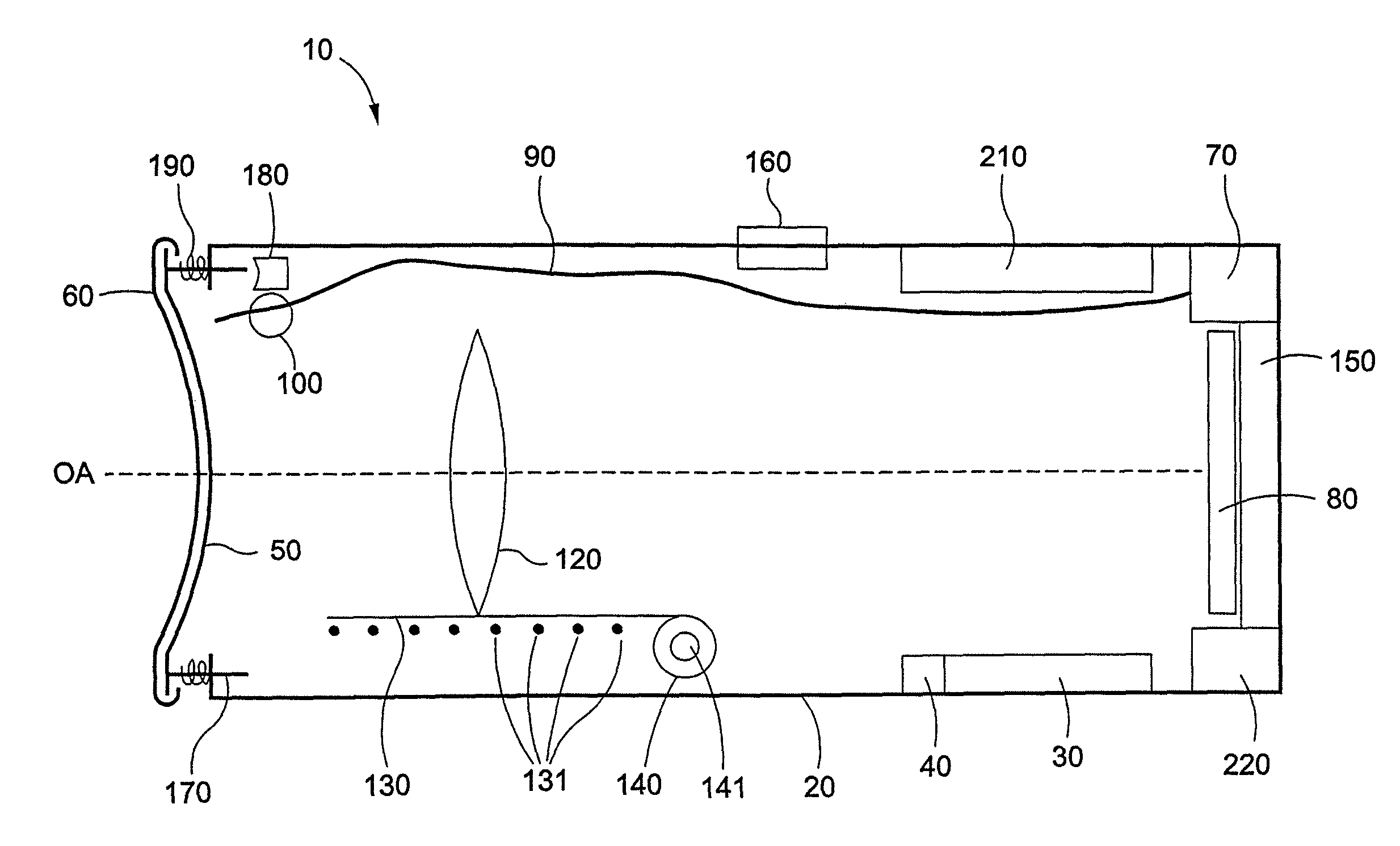
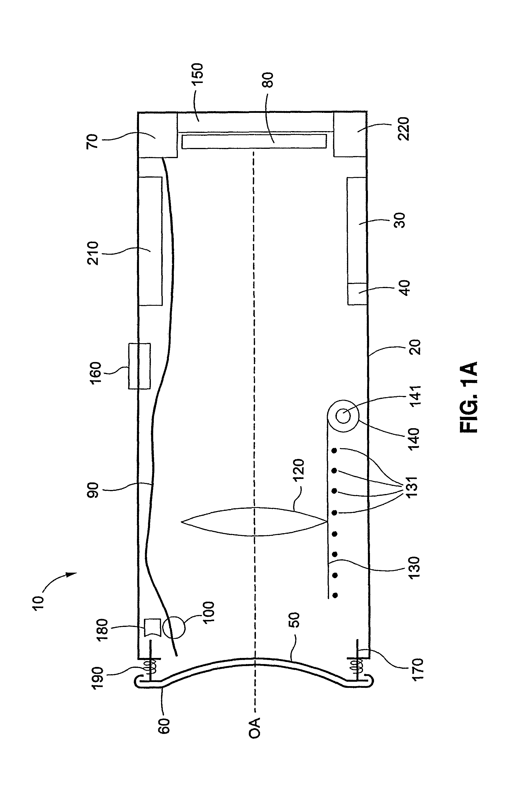
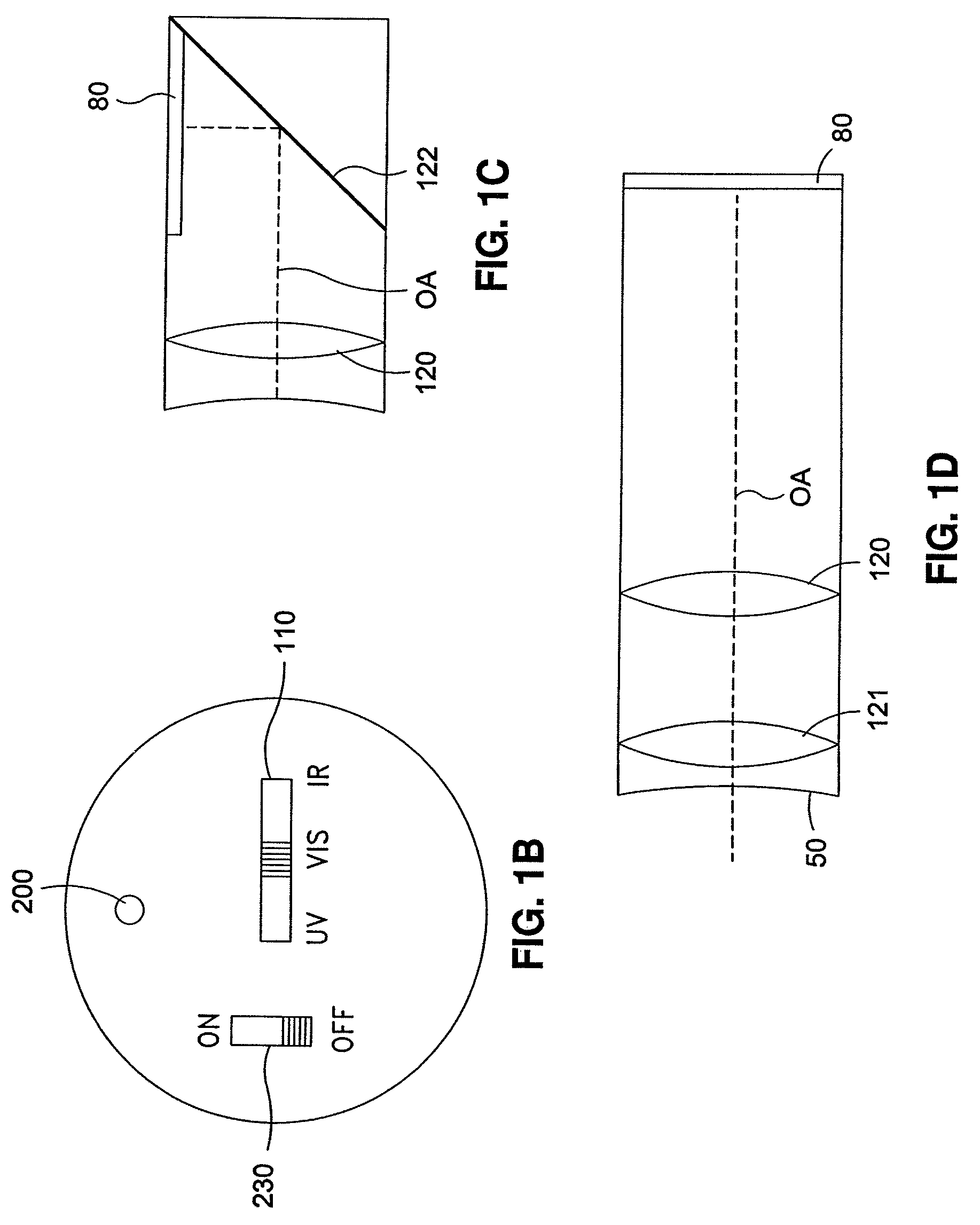
[0058]Embodiments of the present disclosure describe a compact ocular fundus camera that can be both easy to use and fully portable. In general terms, the camera will comprise at least a contact surface adapted to contact the cornea of a patient's eye, an illumination source to illuminate the fundus, an image detector that receives an image of the fundus from light reflected back from the eye and outputs an image data file, and an imaging lens, operative to focus light reflected back from the fundus onto the imaging surface of the image detector. The camera will also comprise communication capability, either wired or wireless to permit transmission of image data to at least one peripheral device. In some embodiments, the camera is simple...


PUM


Abstract

Embodiments of a compact camera for imaging the ocular fundus are described. In some embodiments, the camera is a light, handheld camera that acquires a plurality of images of the fundus. The camera can be configured to acquire images manually or automatically. A movable imaging lens or movable image detector provides the images in a series to obtain an image with optimal focus. The camera can acquire a series of images and allow selection of an optimal image either manually, or automatically. In some embodiments, the camera is part of an imaging system that includes a base station. The base station exchanges data with the camera and can receive image data generated by the image detector of the camera. The base can further include networking capability, such that image data can be distributed over a network or to other communication or computing devices.

Description

RELATED APPLICATIONS[0001]This application claims priority to U.S. provisional application 60 / 847,669, filed Sep. 28, 2006, the entire contents of which are herein incorporated by reference.FIELD OF THE INVENTIONS[0002]Embodiments of the disclosure are useful in the area of medical imaging apparatus, particularly in the area of apparatus for use in imaging a fundus of an eye.BACKGROUND OF THE INVENTIONS[0003]Screening for eye diseases has become increasingly important as the population ages in the United States and around the world. Major causes of irreversible blindness include diabetic retinopathy, macular degeneration, and glaucoma. Standard screening measures such as visual acuity testing or intraocular pressure measurement are inadequate, since these tests only identify patients late in the disease process.[0004]Despite such limitations, there is increasing pressure to screen patients for early indicators of eye disease. For example, health maintenance organizations and governm...

Claims


Application Information

Patent Timeline
Patent Type & Authority Patents(United States)
IPC IPC(8): A61B3/14A61B3/00
CPCA61B3/1208G03B3/02G03B15/03H04N5/2256H04N5/23212G03B2215/0503A61B5/7232A61B5/0013H04N23/56H04N23/661H04N23/676H04N23/63
Inventor FELDON, STEVENYOON, GEUNYOUNG
Owner UNIVERSITY OF ROCHESTER
Why Eureka
Try Eureka