Marker for prolonged rupture of membranes

a technology of amniotic fluid and amniotic membrane, which is applied in the direction of pregnancy proteins, peptide sources, instruments, etc., can solve the problems of amniotic membrane rupture, inability to completely accurately distinguish between amniotic fluid leakage, and difficulty in detecting amniotic fluid leakage, and achieve low levels in maternal serum

Inactive Publication Date: 2011-01-04
SWISS ASIAN PROPERTY
View PDF4 Cites 7 Cited by
  • Summary
  • Abstract
  • Description
  • Claims
  • Application Information

AI Technical Summary

Problems solved by technology

Diagnosis of rupture of the amniotic membranes prior to the onset of uterine contractions remains one of the significant clinical problems in modern obstetrics because there is not a reliable clinical method for the detection of amniotic fluid in the vagina (Gregg, (1992), Obstet. Gynecol. Clin. North Am., 19: p 241; Malee, (1992), Obstet. Gynecol. Clin. North Am., 19: p 309; Zlatnic, (1992), Obstet. Gynecol. Clin. North Am., 19: p 353; Davidson, (1991), Clin. Obstet. Gynecol., 34: p 715).
In other cases PROM is less obvious, and it can be difficult to differentiate between the leakages of amniotic fluid from the presence of urine or endocervical mucus in the vagina.
However, while all of these procedures provide some indication that PROM has taken place they are not completely accurate.
In the most part, previously investigated markers for diagnosing PROM provided false positive results owing to the presence of interfering substances.

Method used


Image

  • Marker for prolonged rupture of membranes
  • Marker for prolonged rupture of membranes
  • Marker for prolonged rupture of membranes

Examples


example 1

Identification and Isolation of PROM Markers

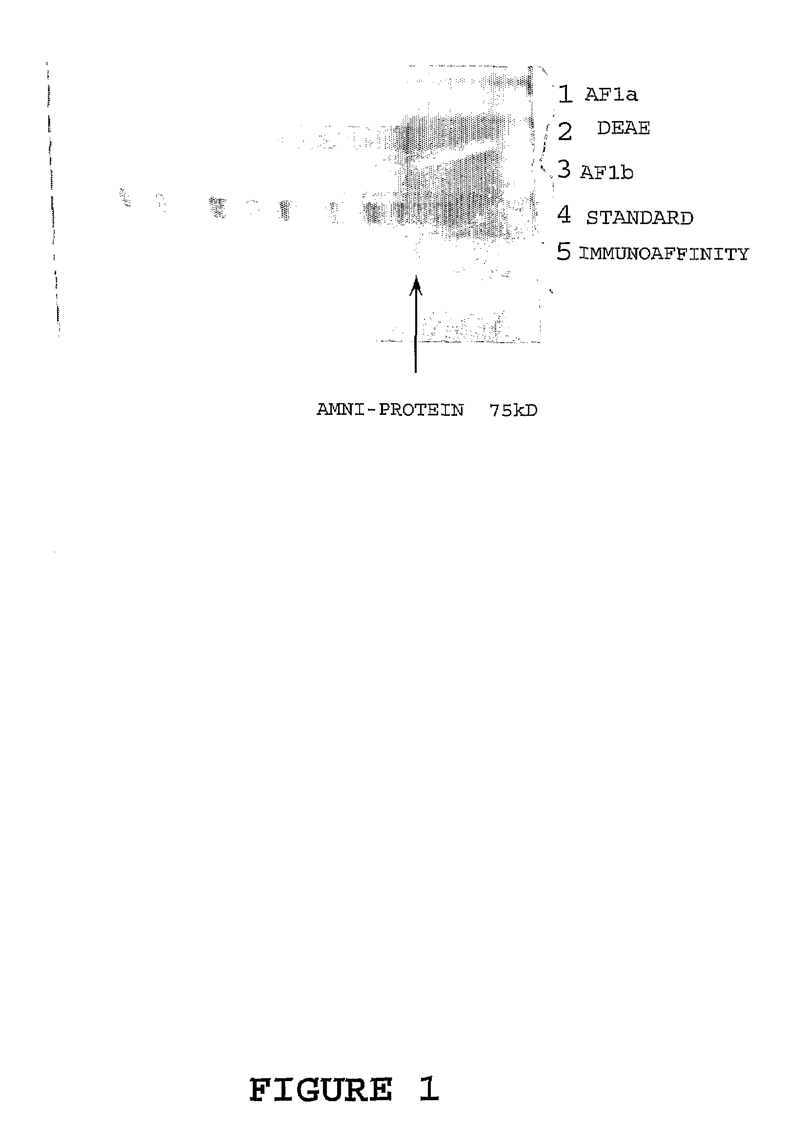
[0109]In the first stage, amniotic fluid specimens were collected (20-975 mL) from women undergoing a caesarean section. All samples were filtered through glass wool prior to being distributed in 2 mL aliquots into sterile 5 mL centrifuge tubes. One tube from each sample was subsequently used in the preparation of various protein fractions (pools) for use in the immunisation schedule and subsequent clinical evaluation.

[0110]Twenty random amniotic fluids of 2 mL were pooled and used in the purification procedure as follows. The entire pool was pretreated with Affi-Gel Blue Gel (AGB [Biorad]) to remove as much of the endogenous albumin as possible. The AGB was prepared by washing with PBS in a 50 mL conical bottom culture tube and centrifuged at 2000 rpm for 10 min. The supernatant was removed and the same volume of fresh buffer was added to the gel, mixed by vortexing, and again centrifuged as above. This procedure was repeated 5 times. AGB...

example 2

Monoclonal Antibody Production

[0115]For primary immunisations of six BALB / c mice, 0.15-0.2 mg of fractions AF1, AF2 and AF3 from Example 1 were emulsified in MPL (1:10 v / v [SIGMA]) and injected subcutaneously into each mouse. Booster immunisations using 0.15-0.2 mg of the respective amniotic fluid fraction prepared with MPL (1:10 v / v [SIGMA]) were given by the same routes. The initial boost was given at three weeks post-immunisation with subsequent boosts prescribed at 7-8 day intervals for 6 weeks. Antibody levels were monitored by collecting eye bleeds 4 weeks post primary immunisation and then weekly until 10 weeks and testing the sera by an indirect one-site enzyme immunoassay.

[0116]In the indirect one-site enzyme immunoassay procedure, 100 μL of each amniotic fluid fraction (0.2 mg / mL) in Tris-HCl buffer pH 7.2, two different amniotic fluid (positive controls) and maternal serum (negative controls) AGB pre-treated pools were added to each well of 96-well microtitre plates [NUNC...

example 3

Indirect 1-Site Screening Enzyme Immunoassay

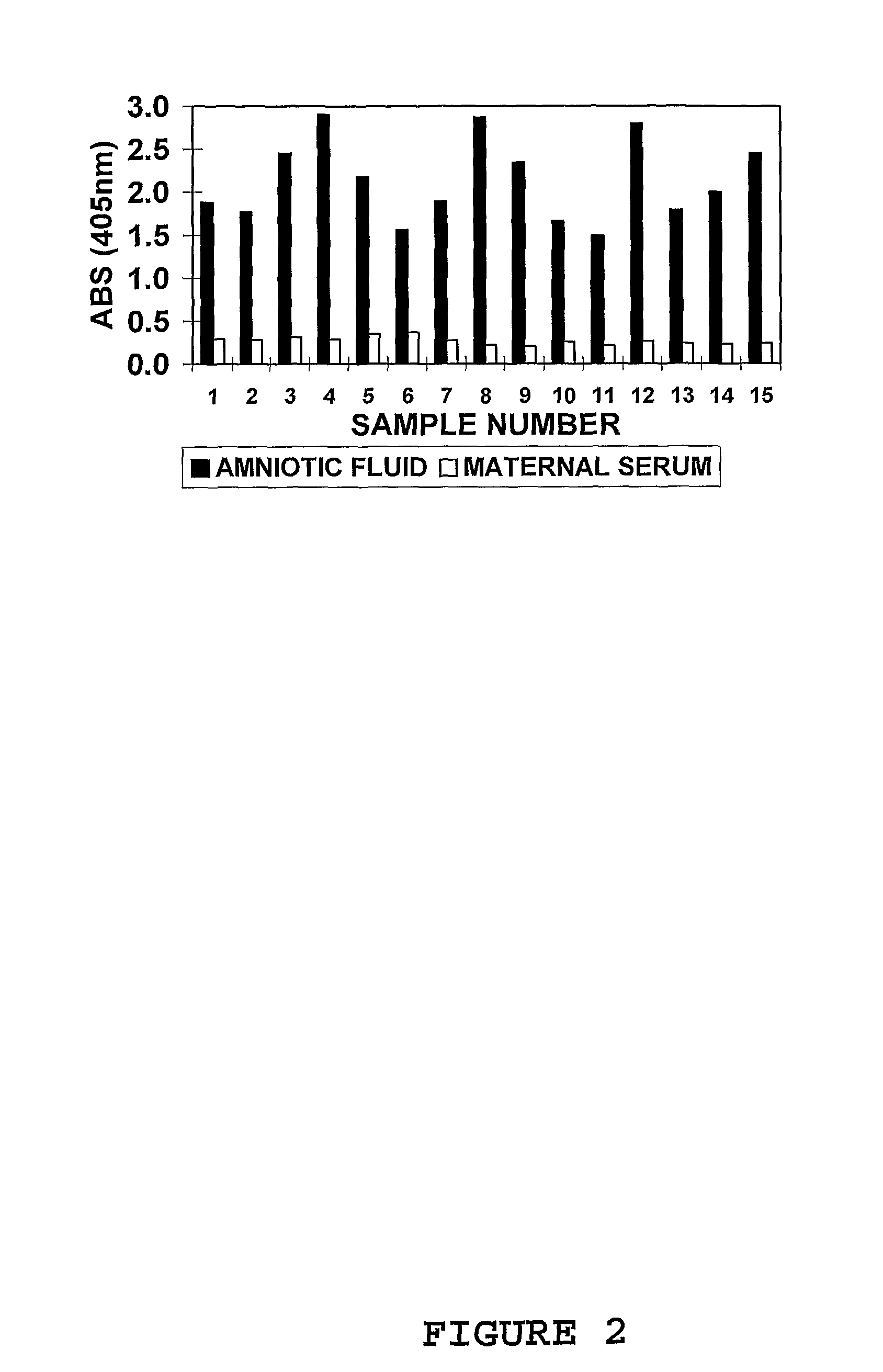
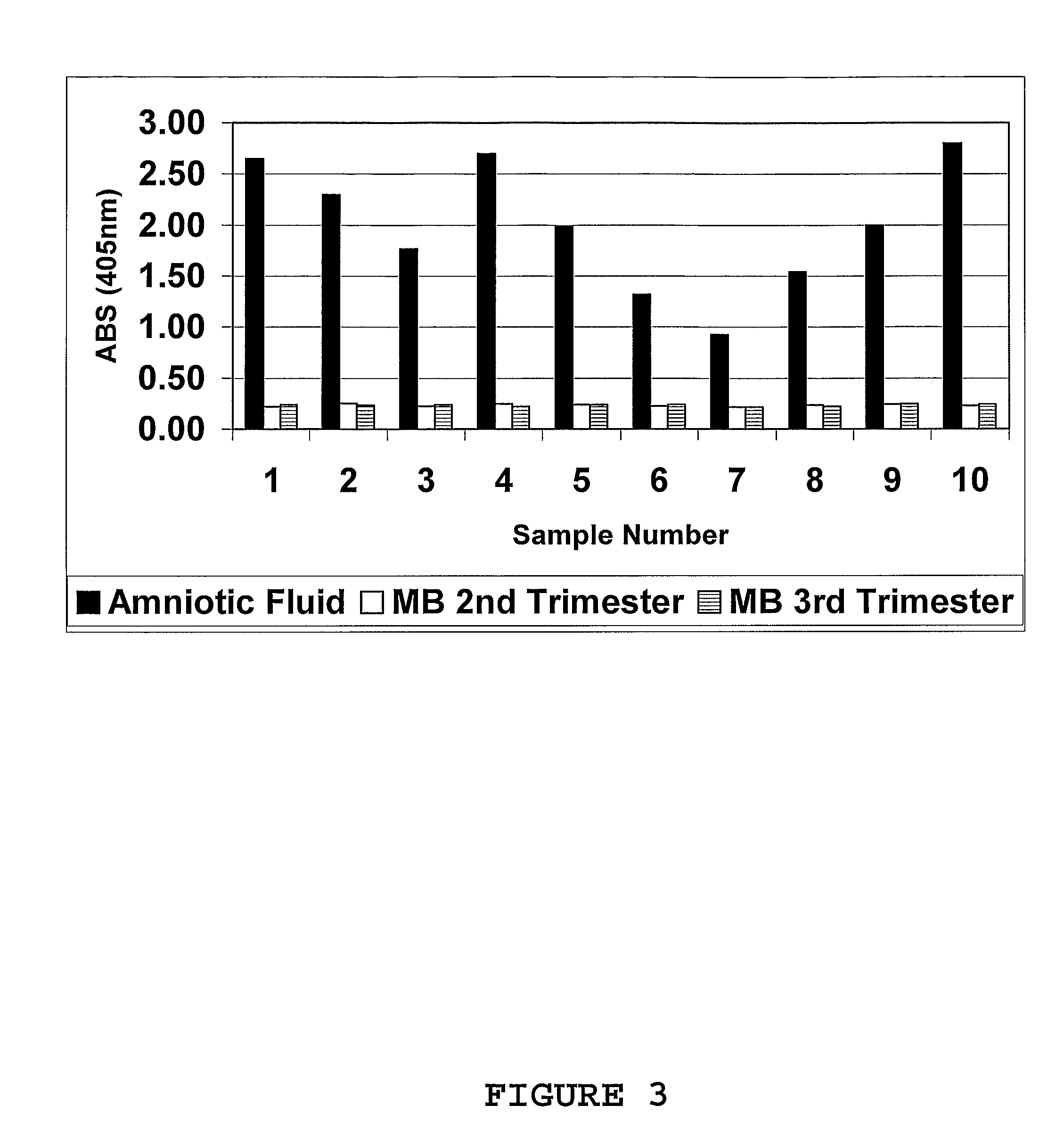
[0123]Amniotic fluid samples (n=6 to 10) and maternal blood (plasma or serum; n=6 to 10) obtained from different patients were pooled to provide a final working volume of 50-100 mL. Sample pools were pre-treated with AGB; 1:1 (v / v) for amniotic fluid and 6:1 (v / v) for maternal blood and mixed end-over-end for 4 hours. The solution was centrifuged at 2000 rpm for 20 min and the supernatants collected into fresh 50 mL sterile culture tubes.

[0124]In the indirect 1-site enzyme immunoassay procedure, 100 μL of both amniotic fluid (positive control / s) and maternal blood (negative controls; plasma or serum obtained from pregnant women during their second and third trimesters) were applied in columns to a 96-well microtitre plate / s (NUNC Immunoplate: maxisorp) and incubated at room temperature (approx. 25° C.) for 1 hour. Two different pools of each respective fluid were used in the immunoassay procedure on the day. Plates were washed in PBS (LS) ...


PUM

PropertyMeasurementUnit
molecular weightaaaaaaaaaa

Abstract

The present invention relates to amniotic fluid specific polypeptide and a method for detecting rupture of the amniotic membranes. In particular the invention provides a marker for prolonged rupture of the membranes (PROM), which marker consists essentially of polypeptides of approximately 75 kDa, 20 kDa and 50 kDa as determined by 4-15% gradient SDS-PAGE under reducing conditions.

Description

FIELD OF THE INVENTION[0001]The present invention relates to amniotic fluid specific polypeptide and a method for detecting rupture of the amniotic membranes. In particular, the present invention relates to a marker for prolonged rupture of the membranes (PROM), which marker consists essentially of polypeptides of approximately 75 kDa, 20 kDa and 50 kDa as determined by 4-15% gradient SDS-PAGE under reducing conditions.BACKGROUND OF THE INVENTION[0002]Diagnosis of rupture of the amniotic membranes prior to the onset of uterine contractions remains one of the significant clinical problems in modern obstetrics because there is not a reliable clinical method for the detection of amniotic fluid in the vagina (Gregg, (1992), Obstet. Gynecol. Clin. North Am., 19: p 241; Malee, (1992), Obstet. Gynecol. Clin. North Am., 19: p 309; Zlatnic, (1992), Obstet. Gynecol. Clin. North Am., 19: p 353; Davidson, (1991), Clin. Obstet. Gynecol., 34: p 715).[0003]Pre-labour rupture of the fetal membranes...

Claims


Application Information

Patent Timeline
Patent Type & Authority Patents(United States)
IPC IPC(8): G01N33/53
CPCC07K14/4715C07K16/18G01N33/689
Inventor VOROTELIAK, VICTOR
Owner SWISS ASIAN PROPERTY
Why Eureka
Try Eureka