Detecting bacteria in fluids

a technology of fluid and detection method, applied in the field of detecting bacteria and optically testing urine, can solve the problems of time and resource consumption, requiring skilled operators, and both above mentioned methods failing to detect bacteria that do not generate those products,

Inactive Publication Date: 2007-09-13
WEISCHSELBAUM AMNON
View PDF4 Cites 21 Cited by
  • Summary
  • Abstract
  • Description
  • Claims
  • Application Information

AI Technical Summary

Problems solved by technology

Common analyses of urine samples involve microscopy and or culturing, require skilled operators and are time and resource consuming.
However, both above mentioned methods fail in detecting bacteria that do not generate those products detectable by the specific reagents included.
The methods are based on a relatively high bacterial concentration in the sample under test and therefore such screening processes are prone to insufficient sensitivity and relatively low specificity.
Furthermore these screening methods are limited to specific populations of patients; they should not be applied for example to infants and or to pregnant women.

Method used


Image

  • Detecting bacteria in fluids
  • Detecting bacteria in fluids
  • Detecting bacteria in fluids

Examples


example 1

[0051] A simulated analysis of an angular scattering distribution was conducted. A synthetic model of urine was made in which 2-4 microns diameter spheres having the same dielectric constant as that of bacteria were included. Salt particles are represented by spheres having a radius that is smaller than one micron and a matching dielectric constant. Calculations are based on the scattering Mie theory [H. C. van de Hulst. “Light scattering by small particles”, John Wiley & Sons publishing, NY, 1957]. Reference is now made to FIG. 5 showing a plot of the simulated angular scattering distribution function of suspensions of the two kinds of particles described above. Curve 101 represents the intensity of light scattered by the suspension of particles representing bacteria. Curve 102 corresponds to the intensity of light scattered by the small particle representing crystalline salt particles in urine. The intensity is shown in a logarithmic scale and is represented in polar coordinates v...

example 2

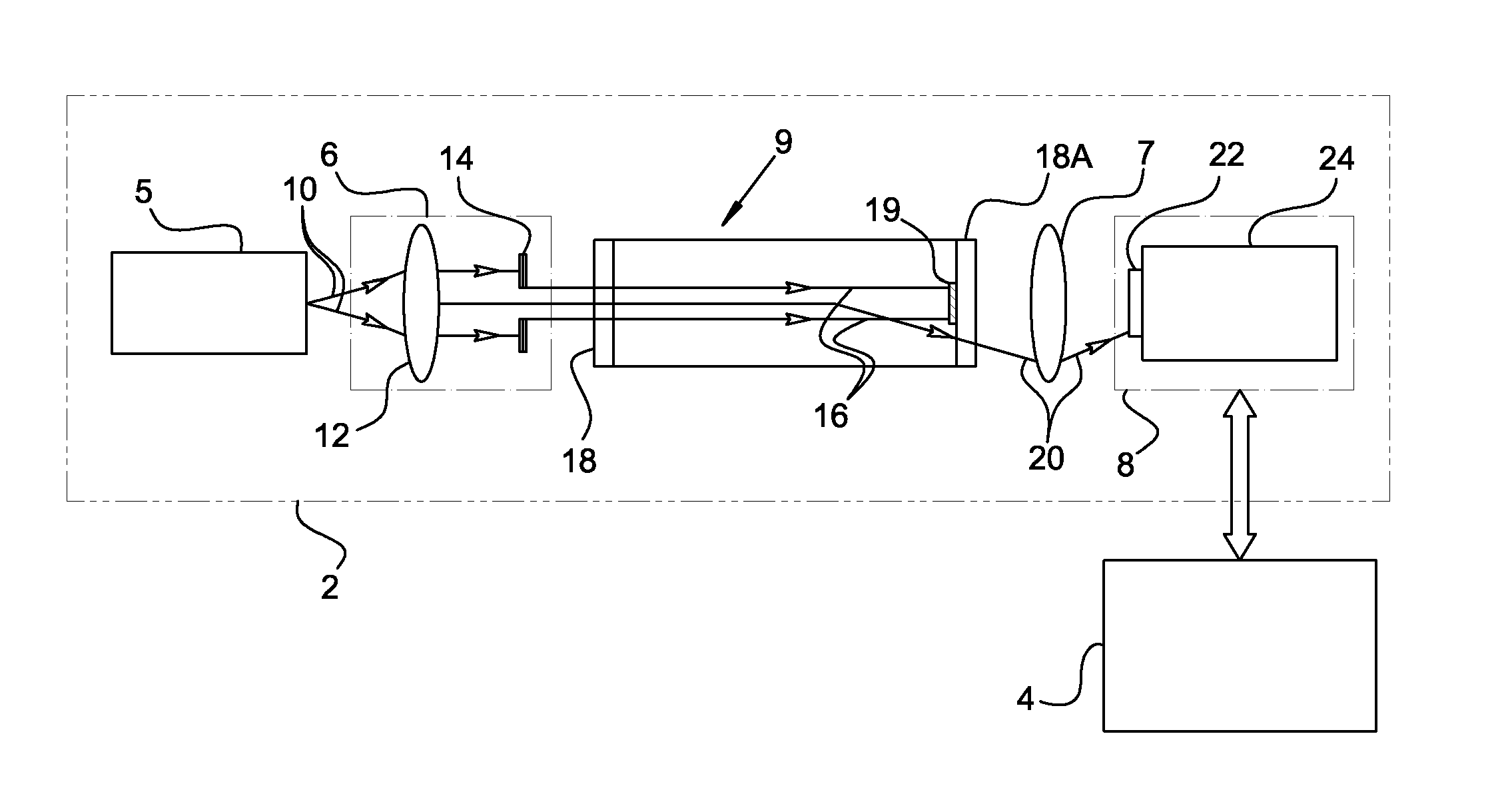
[0052] Measurements described below were taken by employing a system in which the light source is a laser diode of a specific wavelength and intensity and an CU having one cuvette. An elaborated model of urine containing bacteria was prepared and a scaled geometry of the system described in FIG. 1 to which reference is again made was employed as follows: [0053] 1) The dimensions of bacteria are defined in the literature. [0054] 2) The bacteria are randomly dispersed in the illuminated volume of the sample of fluid. [0055] 3) The light source of the model is a laser diode having the same features as the light source of the system employed in the actual measurements. [0056] 4) The angular intensities of scattered light for each single bacterium were calculated using the above mentioned Mie scattering model. [0057] 5) Scattered light rays are optically traced using a model of the optical unit employed for the actual measurements. [0058] 6) The power density of the scattered beam is cal...

example 3

[0061] Light scattering measurements were conducted by employing the same optical unit and CUs as is described in example 2 above and several urine samples supplemented with bacteria at different bacterial concentration levels. Bacterial concentration levels were independently calibrated by employing incubation as in the prior art. Reference is now made to FIGS. 7A-7D in which are shown typical angular scattering distribution functions of infected urine at bacterial concentration levels of 103, 104, 105, and 106 CFU / ml respectively. Scattering profiles were derived from these typical distribution functions by averaging over most of the azimuth range. In FIG. 8 a graph comparing the scattering profiles corresponding to the measured angular distribution functions of FIGS. 7A-7D is shown. Plots 123, 124, 125 and 126 represent these scattering profiles of infected urine at the same bacterial concentration levels as FIGS. 7A-7D respectively.

[0062] A typical urine sample known to be cont...


PUM

PropertyMeasurementUnit
surface roughnessaaaaaaaaaa
widthaaaaaaaaaa
surface roughnessaaaaaaaaaa

Abstract

A method for the detection of bacteria in aqueous fluids in the form of solutions, emulsions and or suspensions, such as drinking water, liquid food and drinks and biological fluids such as urine, spinal and/or amniotic fluids and serums by measurement of light scattering The fluid suspected of containing bacteria is filtered, first to exclude particles of sizes larger than the size of the expected bacteria. Other filtration methods are used in addition to exclude different constituents of the examined fluid. Ion exchange chromatography is such a method. The verification of bacterial contamination and the calculation of its level is performed by matching measured scattering profiles with pre-stored calibrated scattering profiles. A system for carrying out the measurements including cuvette units suited for light scattering measurements is provided.

Description

FIELD OF THE INVENTION [0001] The present invention relates in general to assaying a body fluid. In particular the present invention relates to optically testing urine for the presence of bacteria, light scattering measurements and filtration. BACKGROUND OF THE INVENTION [0002] Aqueous fluids such as solutions, emulsions or suspensions are very common in biological context. These aqueous fluids include potable water for human or animal consumption, liquid food or drinks, urine, amniotic or spinal fluids. Such fluids may be occasionally tested for the presence of bacteria. In clinical microbiology laboratories, a large proportion of analyzed samples are urine samples. Common analyses of urine samples involve microscopy and or culturing, require skilled operators and are time and resource consuming. Therefore, any inexpensive and fast screening method which could obviate a significant amount of expensive and time consuming analytical methods would be beneficial. [0003] Test strips for...

Claims


Application Information

Patent Timeline
Patent Type & Authority Applications(United States)
IPC IPC(8): G01N21/00
CPCC12Q1/04G01N15/0205G01N2021/0321G01N21/51G01N33/493G01N21/0303
Inventor WEISCHSELBAUM, AMNON
Owner WEISCHSELBAUM AMNON
Why Eureka
Try Eureka