Medical imaging system for accurate measurement evaluation of changes in a target lesion

a medical imaging and target lesion technology, applied in the field of medical imaging data analysis, can solve the problems of affecting the clinical trial length, affecting the clinical trial duration, so as to shorten the length of clinical trials

Active Publication Date: 2011-01-25
YANKELEVITZ DAVID DR
View PDF27 Cites 67 Cited by
  • Summary
  • Abstract
  • Description
  • Claims
  • Application Information

AI Technical Summary

Benefits of technology

[0012]In one aspect, the present invention offers a method for shortening the length of clinical trials by providing an accurate method for learning whether a tumor is responding or not in shorter time intervals.
[0013]In another aspect, the present invention offers a method for confidently measuring smaller degrees of change in a tumor to provide information useful for therapeutic response.
[0014]In another aspect, the present invention offers a method for confidently measuring smaller degrees of change in a tumor to provide information useful for diagnostic response.
[0015]In another aspect, the present invention offers a method for confidently measuring smaller degrees of change in a tumor to provide information useful for diagnostic applications.

Problems solved by technology

One of the most expensive and time-consuming aspects of drug development relates to clinical trials for getting anticancer agents such as anticancer drugs approved.
In taking any measurement, accuracy is a critical issue.
Unfortunately, the current RECIST approach for assessment of tumor response to treatment is severely limited because it does not consider measurement accuracy.
As a result, it suffers from the need to observe large changes in single dimensional measurements in order to determine if there has been a response to treatment.
The need for such large changes relates to an inability to reliably make measurements of a tumor size.
Errors related to manually measuring tumor lengths can be quite large.
Similarly, the inability to reliably select comparable imaging planes on temporally separated scans necessitates reliance on large changes in order to be certain that the change is genuine and not one of measurement error.
However, methods for accurate measurement evaluation including, among others, determining a bound on the error of a size change measurement are lacking.

Method used


Image

  • Medical imaging system for accurate measurement evaluation of changes in a target lesion
  • Medical imaging system for accurate measurement evaluation of changes in a target lesion
  • Medical imaging system for accurate measurement evaluation of changes in a target lesion

Examples


Embodiment Construction

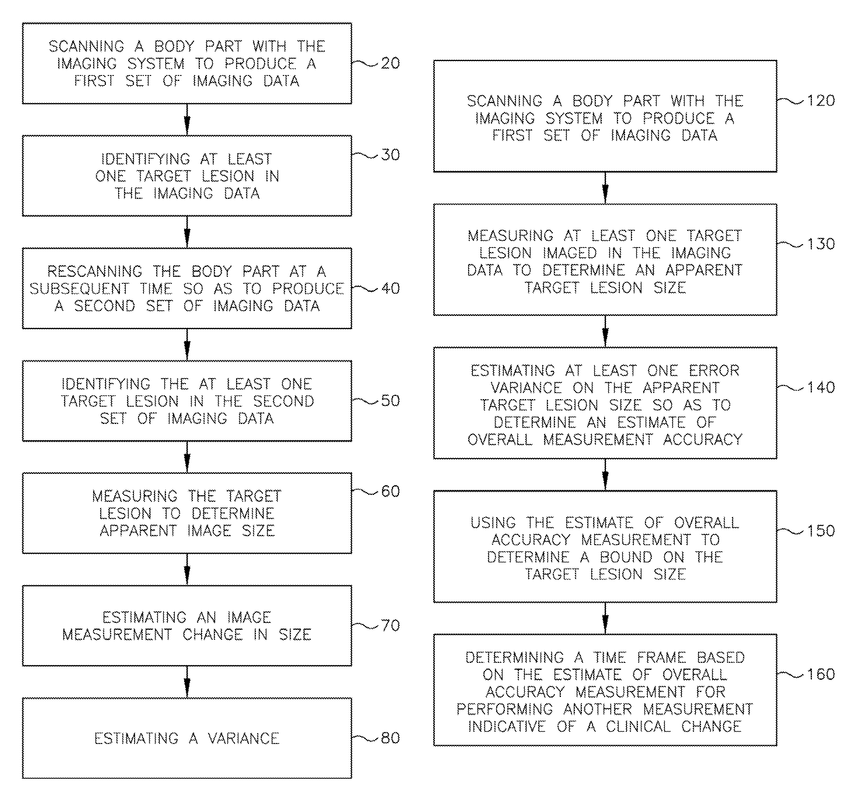
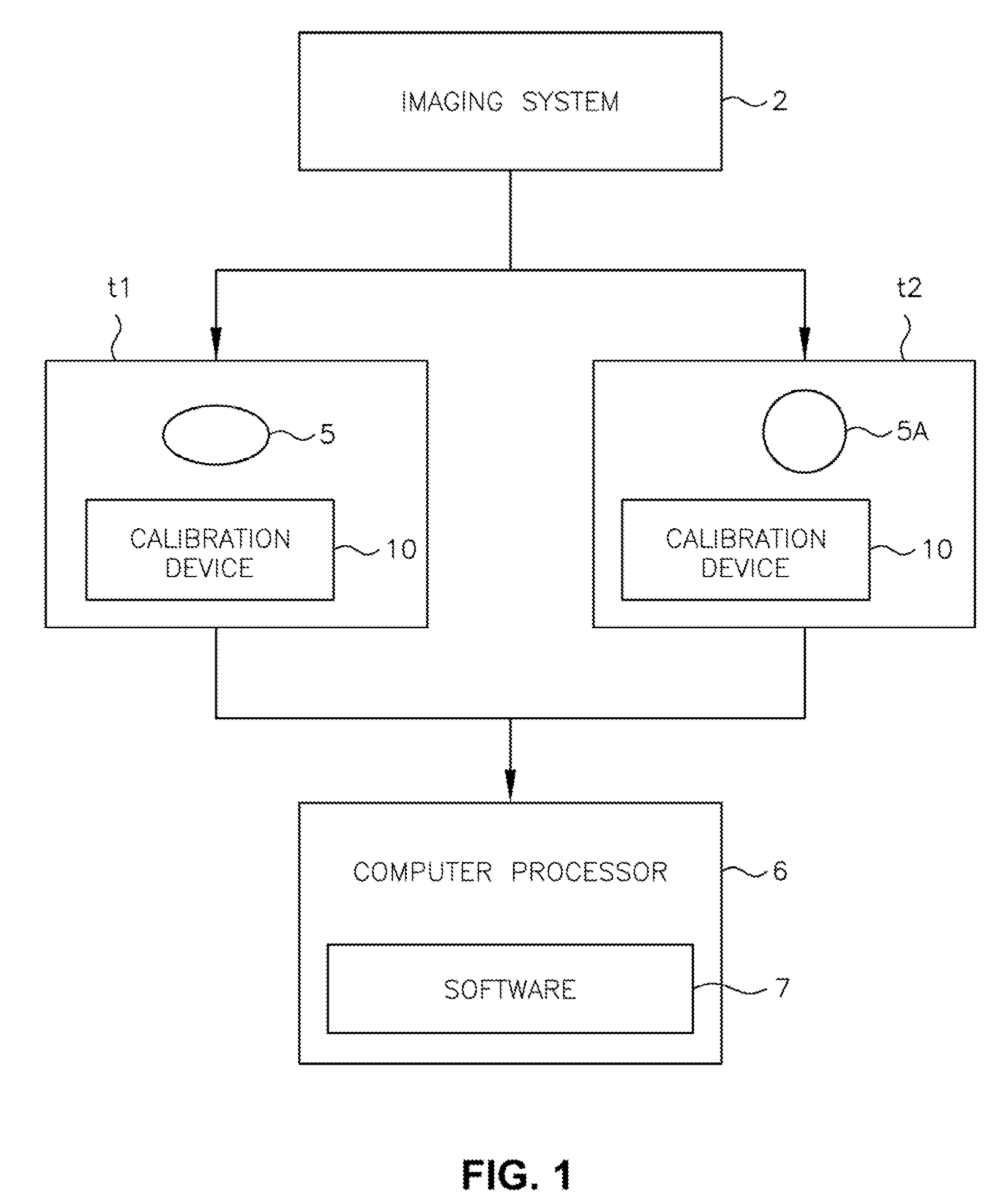
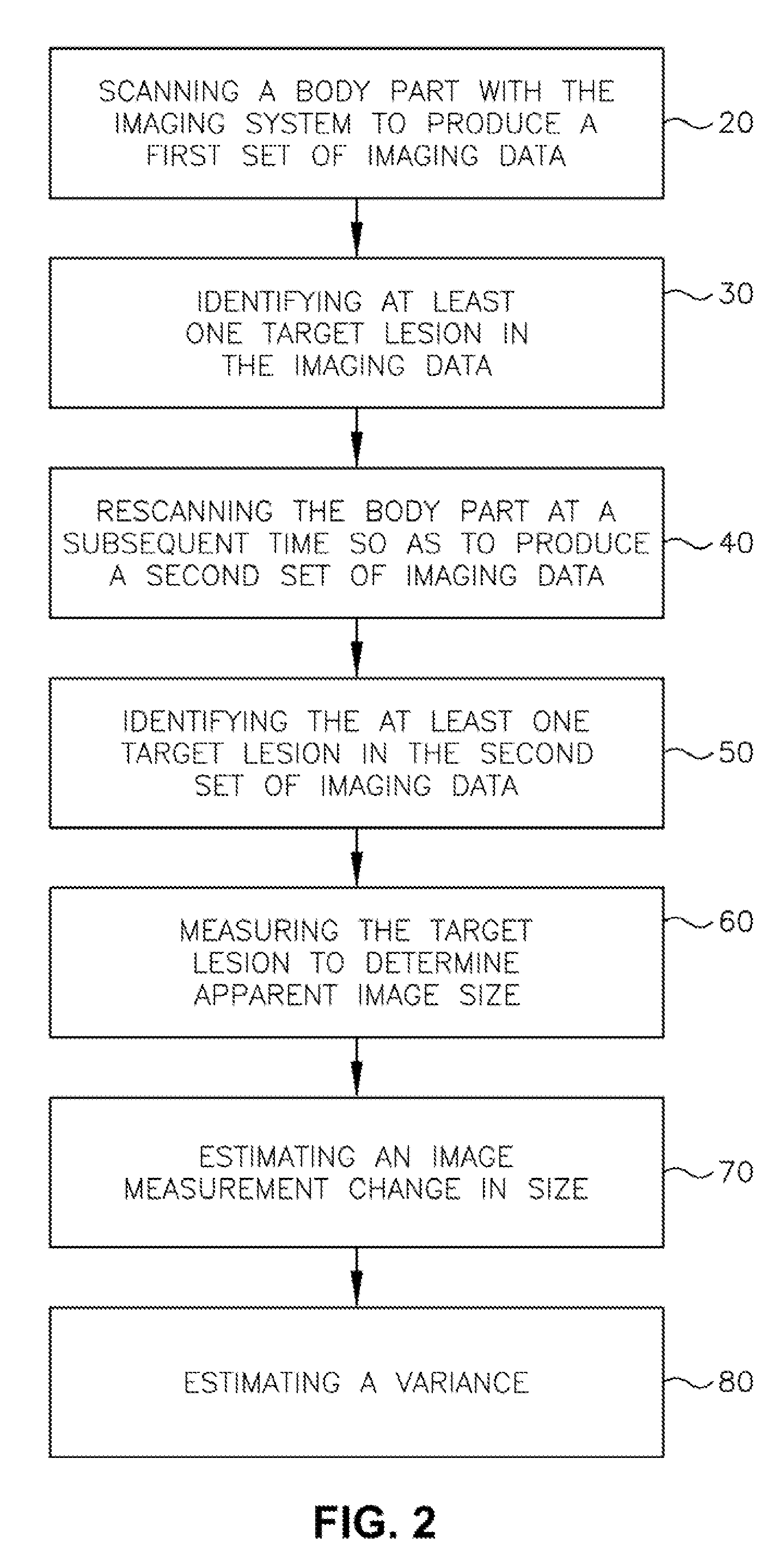
[0031]Preliminarily, it should be noted that, while a particular system and method is described in detail herein for analyzing medical imaging data, such as radiology data, this is not by way of limitation, but solely for the purposes of illustration, and the invention may also be employed for analyzing data of other types.

[0032]The present invention builds on advances in imaging technology that have now made it possible to scan tumors such that the entire tumor volume is imaged. There have been significant improvements in the methods for the measurement of tumor size from CT images over the last decade by using 3D volumetric computer algorithms. In addition, images are not obtained isotropically, meaning that the resolution is nearly the same in the x, y, and z dimensions. Advanced image processing allows for improved segmentation of the tumor from surrounding structures, with better definition of the tumor boundaries, thus leading to improved measurements.

[0033]The present inventi...


PUM


Abstract

A body part is scanned to produce a first set of imaging data. A target lesion in the imaging data is identified. The body part is rescanned at a subsequent time so as to produce a second set of imaging data. The target lesion is identified in the second set of imaging data and the size of the target lesion is measured in the first and second sets of imaging data to determine two apparent image volumes corresponding to the first and second sets of imaging data. A change in size is estimated by comparing the first and second apparent lesion sizes. A variance on the change in size is estimated so as to determine a bound on the change in size measurement.

Description

RELATED APPLICATIONS[0001]This application claims priority from co-pending international application number PCT / US2005 / 013968, entitled MEDICAL IMAGING SYSTEM FOR ACCURATE MEASUREMENT EVALUATION OF CHANGES IN A TARGET LESION, having international filing date Apr. 25, 2005, which, in turn claims priority from U.S. Provisional application Ser. No. 60 / 565,327, of the same title, filed Apr. 26, 2004, both applications are incorporated by reference.FIELD OF THE INVENTION[0002]The present invention relates generally to analysis of medical imaging data, and, more particularly to an automated computer process for accurate measurement evaluation of changes in a target lesion or multiple target lesion as imaged by a medical imaging system.BACKGROUND OF THE INVENTION[0003]The pharmaceutical industry develops a variety of products that require approval from the FDA often based on measurements derived from medical images. One of the most expensive and time-consuming aspects of drug development r...

Claims


Application Information

Patent Timeline
Patent Type & Authority Patents(United States)
IPC IPC(8): G06K9/00A61B5/05A61B6/00A61B6/12G06T7/00
CPCA61B5/1075A61B6/032A61B6/583G06T7/0012A61B6/12G06T2207/30004G06T7/12A61B6/03G06T11/60G06T2207/30096
Inventor YANKELEVITZ, DAVID F.REEVES, ANTHONY P.HENSCHKE, CLAUDIA INGRID
Owner YANKELEVITZ DAVID DR
Why Eureka
Try Eureka