Cathepsin E as a marker of colon cancer

a marker and cathepsin technology, applied in the field of colon cancer detection methods, can solve the problems that the detection of tissue or urine cate levels has not yet been established for the diagnosis or detection of colon cancer, and the non-invasive test, such as the urine test, has not yet been established for colon cancer diagnosis or detection, and achieves the effect of non-invasive and readily implemented

Inactive Publication Date: 2011-02-22
UNIV OF NOTRE DAME DU LAC
View PDF4 Cites 2 Cited by
  • Summary
  • Abstract
  • Description
  • Claims
  • Application Information

AI Technical Summary

Benefits of technology

[0016]In another aspect of the present invention, a method for diagnosing and/or providing a routine clinical screen for colon cancer is provided. In some embodiments, the method comprises measuring a cathepsin E concentration in a test urine sample to provide a test cathepsin E concentration, and comparing the test cathepsin E concentration to

Problems solved by technology

However, a detectable increase in tissue or urine catE levels has not been established for the diagnosis or det

Method used


Image

  • Cathepsin E as a marker of colon cancer
  • Cathepsin E as a marker of colon cancer
  • Cathepsin E as a marker of colon cancer

Examples


example i

Materials and Methods

[0031]The present example sets forth the various materials and methods that were used in generating the disclosure presented herein.

Mice and Tissue Processing

[0032]Mice were maintained in a facility accredited by the Association for Assessment and Accreditation of Laboratory Animal Care (AAALAC), International. Mouse protocols were reviewed and approved by the University of Notre Dame Institutional Animal Care and Use Committee. The APCMin / + colony was generated from C57B16 / J APCMin / + mice purchased from Jackson Laboratories (Bar Harbor, Me.). The mice were kept on a standard rodent diet. Animals were sacrificed by CO2 asphyxiation, and their intestines removed, rinsed in phosphate-buffered saline, and incised. Tissues were immediately fixed with 10% formalin, processed, embedded in paraffin, and sectioned (4 μm). Sections were stained with hematoxylin-eosin (H & E).

Laser Capture Microdissection (LCM)

[0033]Cryosections (8 μm) from the frozen ilea of 90-day old m...

example 2

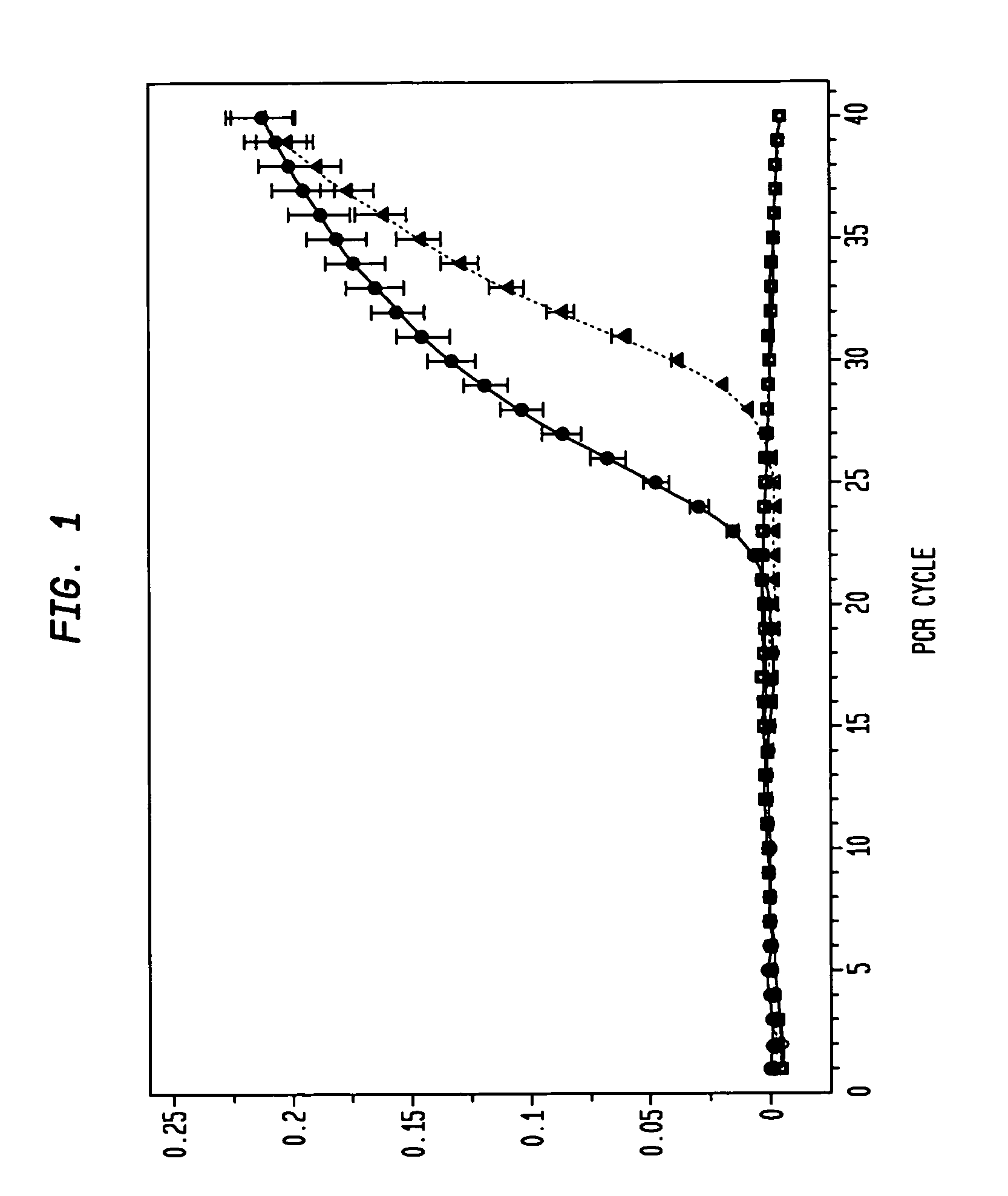
Real-Time RT-PCR Detection of catE mRNA Expression in APCMin / + Adenomas

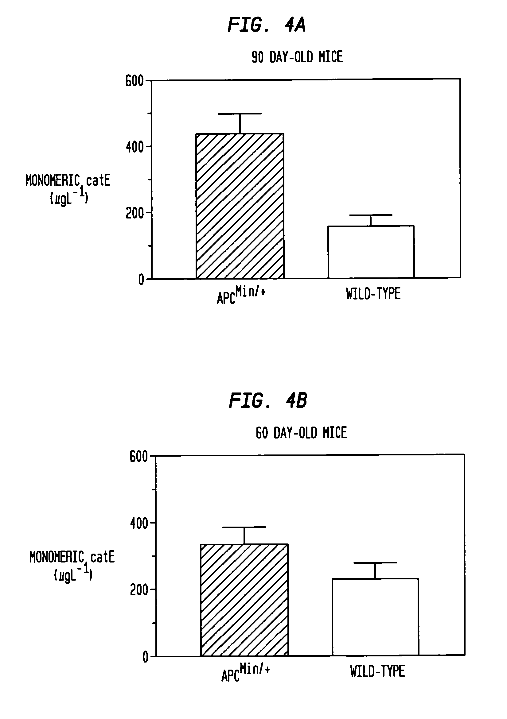
[0043]The present example demonstrates the utility of the invention for providing an RNA profiling screening method for diagnosing and / detecting and monitoring the progression of colon cancer. In this regard, the present example demonstrates that an increase in catE mRNA transcription, and catE expression (monomeric catE) is markedly increased in dysplastic cells, but not in normal (i.e., from non-dysplastic tissue) cells.

[0044]To ensure a homogeneous population of cell types for RNA isolation, LCM was employed for the selection of intestinal epithelia corresponding to adenomatous APCMin / +, normal APCMin / +, and normal wild-type tissue. Total mRNA from wild-type spleen, in which abundant levels of catE transcript have been previously established [7], was employed as an exogenous positive control.

[0045]The results of the real-time RT-PCR monitoring are presented in FIG. 1. After 40 PCR cycles, no amplification of c...

example 3

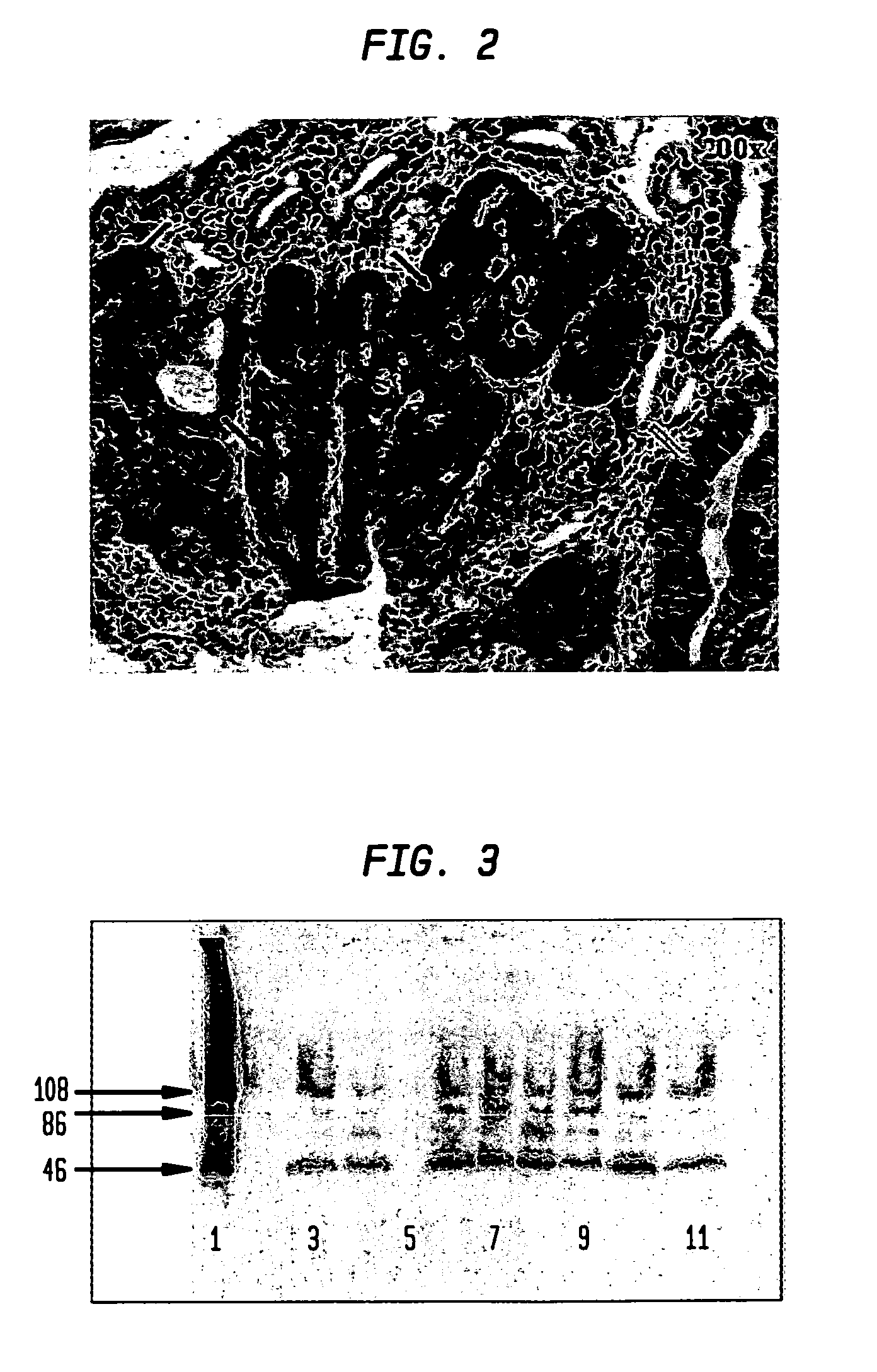
Immunohistochemical Detection of catE in Intestinal Lesions of APC Min / + Mice

[0046]The present example demonstrates that dysplastic tissue is immunohistochemically detectable by the presence of specifically staining tissue using an anti-catE antibody, and that the intensity of immunohistochemical staining obtained is correlated to the severity of the dysplastic lesion.

[0047]Intestinal sections from a total of 33 90-day old APCMin / + mice were subjected to immunohistochemical analyses with a commercially available polyclonal antibody to catE. Examined sections manifested gastrointestinal intraepithelial neoplasia (GIN) lesions and adenomas; no adenocarcinomas were observed. A representational section of an APCMin / + mouse intestine (FIG. 2) shows catE staining in all regions of the epithelia manifesting mild to severe dysplasia. Normal-appearing areas of the intestine are devoid of detectable staining, as was also observed for sections from wild-type mice. A direct relationship is show...


PUM

PropertyMeasurementUnit
Fractionaaaaaaaaaa
Atomic weightaaaaaaaaaa
Concentrationaaaaaaaaaa

Abstract

Elevated levels of cathepsin E (catE) are demonstrated to be diagnostic of intestinal forms of cancer, such as colorectal cancer. Elevated levels of cathepsin E (catE, monomeric forms) are demonstrated to be detectable in the urine of animals having colorectal cancer, and a diagnostic/screening method for identifying and/or detecting colorectal in an animal from a urine sample is provided. Specific tissue immunohistochemcial staining for catE (monomeric forms) in dysplastic tissue is also disclosed, and is shown to correlate with the level of dysplastic lesion severity. Hence, a method for determining and identifying dysplastic lesion severity is provided. Cathepsin E mRNA transcription and expression levels are also demonstrated to be upregulated in dysplastic tissue, relative to non-dysplastic tissue. Hence, a method for transcriptionally profiling an animal to monitor the progression of colorectal disease is provided.

Description

CROSS-REFERENCE TO RELATED APPLICATIONS[0001]This application claims priority to U.S. Provisional App. No. 60 / 638,240, entitled “Cathepsin E as a Marker of Colon Cancer,” filed Dec. 23, 2004. The entire disclosure and contents of the above application is hereby incorporated by reference herein.GOVERNMENT INTEREST STATEMENT[0002]The United States Government has rights in this invention pursuant to federal funding provided in United States Department of Defense Contract No. DAMD17-03-1-0206.BACKGROUND[0003]1. Field of the Invention[0004]The present invention relates generally to methods of detecting colon cancer, and more particularly, to a method for detecting colon cancer using a marker, cathepsin E.[0005]2. Related Art[0006]The identification of molecular markers and proteomic patterns in the inception and progression of colon cancer is a major goal in the management of this disease. In the search for sensitive and accurate markers of colon cancer, APCMin / + mice (heterozygous for a...

Claims


Application Information

Patent Timeline
IPC IPC(8): G01N33/53
CPCG01N33/573G01N33/57419G01N2333/948
Inventor NAVARI, RUDOLPH M.PROROK, MARYCASTELLINO, FRANCIS J.
Owner UNIV OF NOTRE DAME DU LAC
Why Eureka
Try Eureka