Ultrasound energy driven intraventricular catheter to treat ischemia

a technology of ischemia and intraventricular catheter, which is applied in the field of ischemia treatment, can solve the problems of affecting the quality of life of millions of people, excessive trauma and damage to the heart tissue, and the risk of complications such as hemorrhaging and scarring, and achieves the effect of reducing the risk to the patient and the injury to the heart tissu

Inactive Publication Date: 2011-03-08
ABBOTT CARDIOVASCULAR
View PDF50 Cites 99 Cited by
  • Summary
  • Abstract
  • Description
  • Claims
  • Application Information

AI Technical Summary

Benefits of technology

[0016]Embodiments of the present invention include methods and apparatuses for treating ischemic myocardium. The invention minimizes injury to the heart tissue and risk to the patient while still causing the cellular damage believed necessary for revascularization of ischemic tissue.
[0017]In one embodiment, an ultrasonic device is used to form localized, precisely placed thermal lesions in and near the ischemic tissue. Ultrasonic energy can be advantageously locally directed and is relatively easy to control. Therefore, the thermal lesions formed by ultrasonic energy can induce angiogenesis in myocardium without creating channels and without excessive damage and trauma to the tissue. Even beyond inducing angiogenesis, it is believed that the method will assist in increasing blood flow to the treated region and mitigates the progression and symptoms of ischemia.
[0020]An embodiment of an ultrasonic catheter within the present invention can have an array of ultrasonic transducers. Ultrasonic energy at a frequency greater than approximately 1 MHz can be applied from the ultrasonic transducers in the array to create additional thermal lesions. The ultrasonic transducers of the array may be independently coupled to a power source and independently controlled by a controller, allowing ultrasonic energy of varying power and duration to be independently applied from each transducer in the array. The controller can control the duty cycle of the power source, so that higher powers can be applied to the tissue without overheating the transducer. The independently controlled transducers in the array can advantageously be used to create multiple thermal lesions that have a gradient of sizes.
[0021]The ultrasonic transducer can have a shape that causes the ultrasonic energy emitted by the transducer to converge in a region located internal to the myocardium and at a distance from the endocardium and epicardium. The transducer can have, for example, a bowl-like, partial cylinder, or hollow hyperboloid-like shape. Accordingly, the thermal lesions produced by the converging ultrasonic energy will be located internal to the myocardium and distal from the endocardium and epicardium. This allows ischemic region internal to the myocardium to be treated without injuring the endocardium or epicardium.

Problems solved by technology

Heart disease is a significant health problem and impairs the quality of life for millions of people.
A problem with the above procedures is that creating channels in the myocardium causes excessive trauma and damage to the heart tissue.
The epicardium, endocardium, or both are punctured to form the channels, leading to a risk of complications such as hemorrhaging and scarring.
The possibility that an embolus will form and cause, for instance, a stroke is another potential complication with the procedures.
As to problems with the particular methods described above, laser energy is known to kill healthy cells, which may worsen the patient's condition.
The laser procedure may also cause denervation, which relieves the chest pain associated with ischemia, but permanently damages the heart muscle.
In addition, controlling the location and depth of a channel formed by laser or RF energy is difficult, making accidental damage to healthy tissue more likely.
RF energy is also diffuse, making it especially difficult to localize damage from the RF energy device, and creating problems such as the coagulation of surrounding blood.
However, although massaging the tissue is less traumatic to the heart tissue than creating channels, massaging alone does not fully treat ischemia and does not cause the cellular damage necessary to stimulate angiogenesis.

Method used


Image

  • Ultrasound energy driven intraventricular catheter to treat ischemia
  • Ultrasound energy driven intraventricular catheter to treat ischemia
  • Ultrasound energy driven intraventricular catheter to treat ischemia

Examples


Embodiment Construction

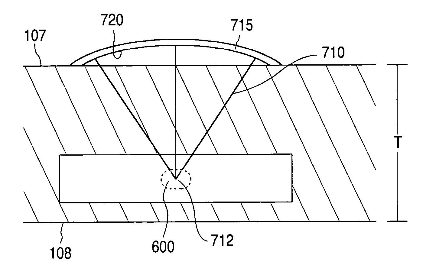
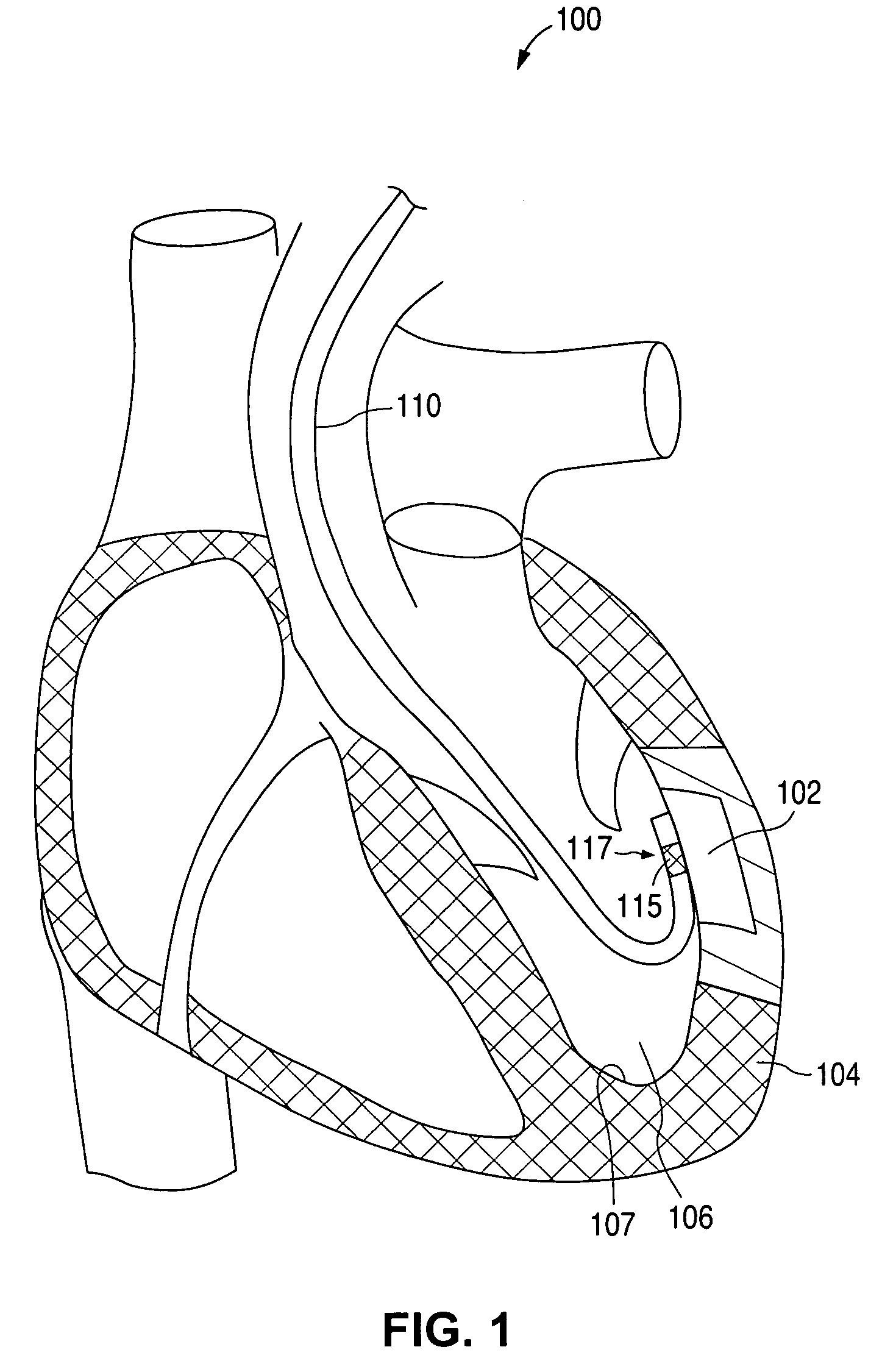
[0035]FIG. 1 illustrates a heart 100 having an ischemic region 102 in the myocardium 104 of the left ventricle 106. A catheter 110 has been inserted into the left ventricle 106. An ultrasound transducer 115 is mounted on distal portion 117 of catheter 110. Ultrasound transducer 115 is positioned adjacent to the endocardial surface 107 and proximate to ischemic region 102, where it is used to treat ischemic region 102, as described below. In particular, transducer 115 is positioned on the endocardial surface 107 laterally adjacent ischemic region 102.

[0036]Although FIG. 1 illustrates ischemic region 102 and catheter 110 in the left ventricle 106, which is particularly susceptible to ischemia, the beneficial effect of the procedures and devices described herein can be used to treat any ischemic area of the heart or other body tissue.

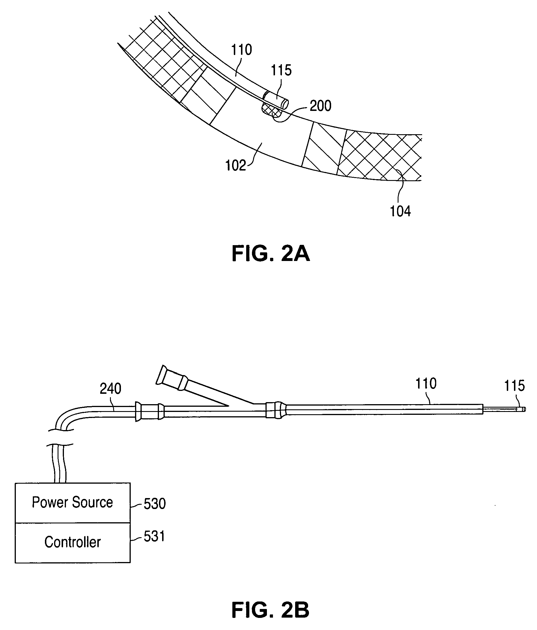
[0037]FIG. 2A illustrates a portion of myocardium 104 with an ischemic region 102. Ultrasound transducer 115 on catheter 110 is oriented toward ischemic r...


PUM


Abstract

A method and apparatus for improving blood flow to an ischemic region (e.g., myocardial ischemia) a patient is provided. An ultrasonic transducer is positioned proximate to the ischemic region. Ultrasonic energy is applied at a frequency at or above 1 MHz to create one or more thermal lesions in the ischemic region of the myocardium. The thermal lesions can have a gradient of sizes. The ultrasound transducer can have a curved shape so that ultrasound energy emitted by the transducer converges to a site within the myocardium, to create a thermal lesion without injuring the epicardium or endocardium.

Description

CROSS REFERENCE[0001]This application is a continuation of of U.S. patent application Ser. No. 10 / 817,358, now U.S. Pat. No. 7,001,336 filed on Apr. 1, 2004, which is a continuation of U.S. patent application Ser. No. 09 / 774,334, now U.S. Pat. No. 6,740,040 filed on Jan. 30, 2001.BACKGROUND[0002]1. Field of the Invention[0003]The invention relates generally to treatment of heart disease. More particularly, the invention is directed to an ultrasonic method and apparatus to treat ischemic tissue.[0004]2. Description of the Background[0005]Heart disease is a significant health problem and impairs the quality of life for millions of people. A common form of heart disease is ischemic heart disease, a condition in which parts of the heart muscle, or myocardium, do not receive an adequate supply of blood. Typically, this condition occurs when the arteries that carry blood to the myocardium become clogged by plaque build-up on their inner walls. The clogged arteries hinder blood flow, and t...

Claims


Application Information

Patent Timeline
Patent Type & Authority Patents(United States)
IPC IPC(8): A61B17/00A61N7/02
CPCA61N7/022A61B2017/00247A61B2018/00392
Inventor MANDRUSOV, EVGENIASTEWARD, JEFFREYSHERMAN, MARSHALL
Owner ABBOTT CARDIOVASCULAR
Why Eureka
Try Eureka