Method and apparatus for classification of coronary artery image data

a technology of image data and classification method, applied in image enhancement, medical/anatomical pattern recognition, instruments, etc., can solve the problems of requiring a lot of computation time and power, and achieve the effect of reducing the effect of any anatomical peculiarities

Inactive Publication Date: 2011-05-10
TOSHIBA MEDICAL VISUALIZATION SYST EURO
View PDF2 Cites 24 Cited by
  • Summary
  • Abstract
  • Description
  • Claims
  • Application Information

AI Technical Summary

Benefits of technology

[0007]This method provides an accurate and computationally efficient technique for automatically classifying and labeling coronary artery image data. A correctly labeled image can thereby be presented to a user or a correctly classified data set can be exported to diagnostics software with little or no need for intervention by a clinician or other medical staff. Typically only a quick verification of the correctness of the labels is required, and in most cases no or very few changes are needed. This can increase the speed of handling if a large number of patient images are required to be processed.
[0009]A benefit of using topological and geometric rules for candidate elimination is that the relevant features defined by these rule sets can be identified from a simple polyline tree representation of the full three-dimensional arterial tree image. Thus the classification processing can be conducted using a much smaller quantity of data than the alternative of handling the full image volume data set or even a segmented version of the arterial tree image. Thus, the computational cost is further reduced.
[0016]The predetermined mean feature vectors may be the mean values of a plurality of feature vectors calculated for correctly classified vessels in arterial trees imaged in other data sets, and the variance-covariance matrices may be calculated for correctly classified vessels in arterial trees imaged in the other data sets. This mitigates the effects of any anatomical peculiarities or errors in the images which could otherwise adversely affect the feature comparison. The use of mean values obtained from a plurality of data sets in effect produces standardised feature vectors reflecting “average” arterial vessel structures.

Problems solved by technology

To consider every feasible classification in detail to identify the correct classification would require a lot of computation time and power.

Method used


Image

  • Method and apparatus for classification of coronary artery image data
  • Method and apparatus for classification of coronary artery image data
  • Method and apparatus for classification of coronary artery image data

Examples


Embodiment Construction

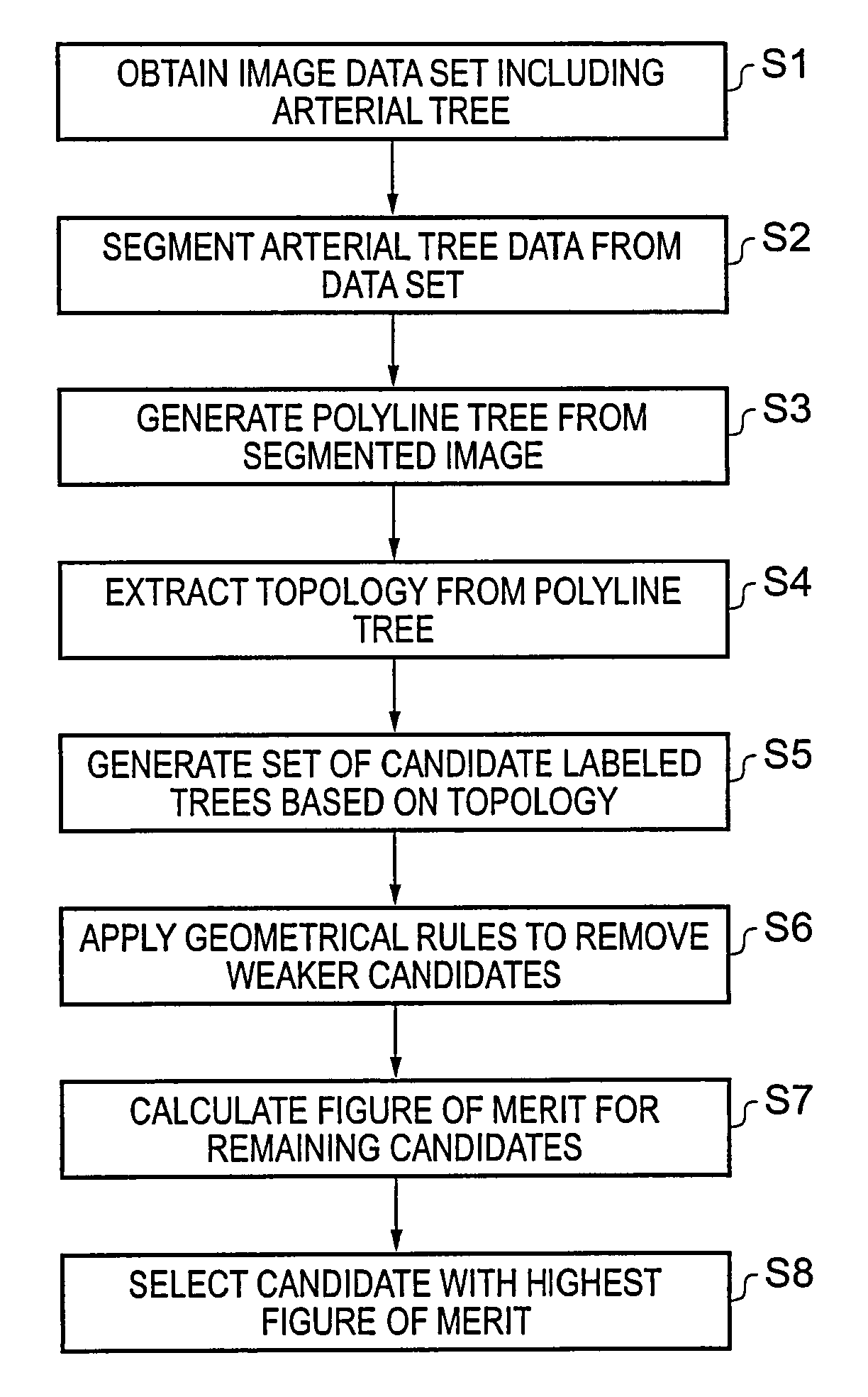
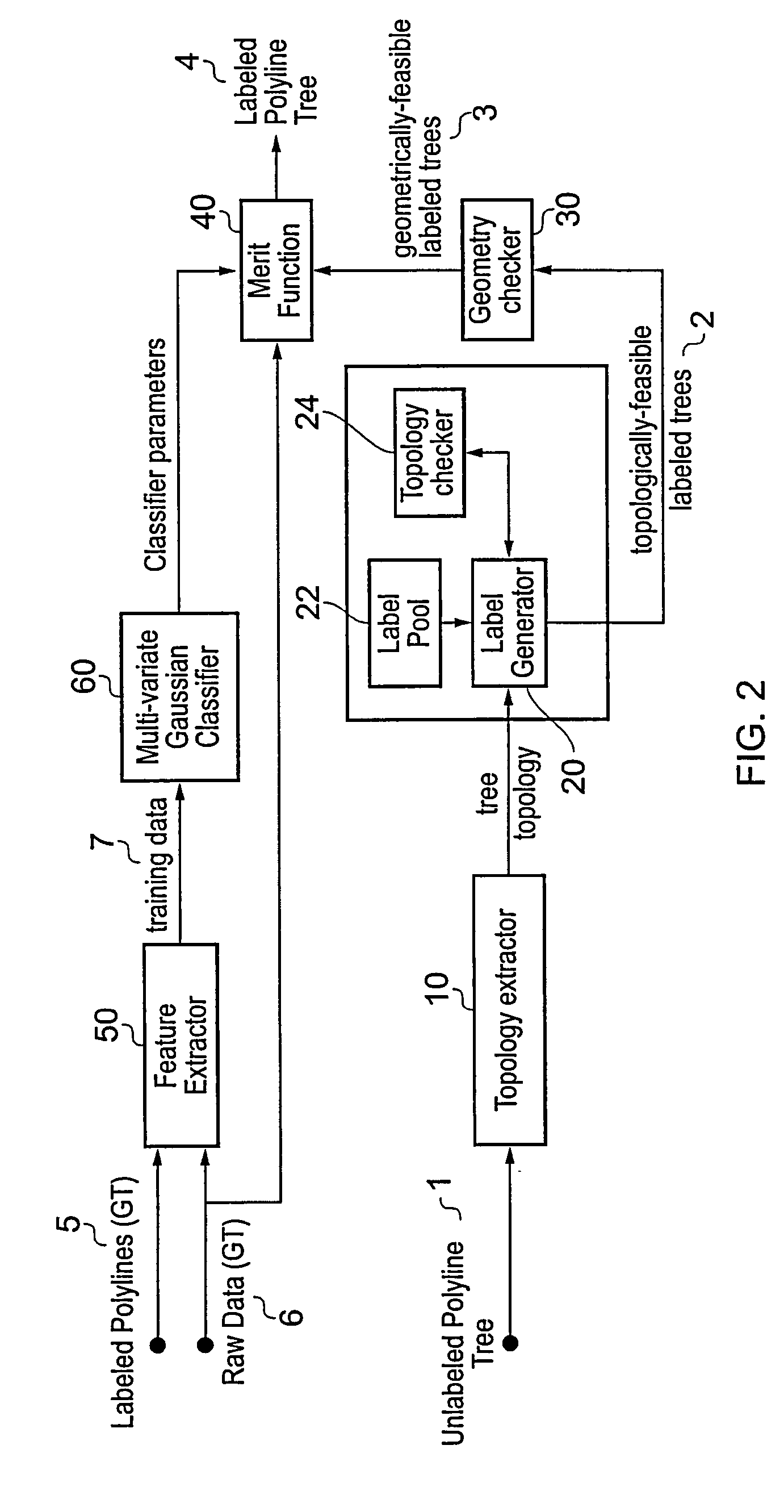
[0029]The human coronary arterial tree comprises the left and right main coronary arteries which branch off from the ascending aorta, and a number of successively smaller vessels which branch off from the main arteries. The present invention seeks to provide a technique for automatic classification and labeling of the various vessels included in a three-dimensional image of the arterial structure. The technique uses filtering based on topological and geometrical parameters to obtain a set of potentially correctly labeled candidates for an imaged arterial tree. A figure of merit is calculated for each member of the set, and the candidate with the best figure of merit is output as the final labeled tree.

[0030]FIG. 1 shows a flow chart indicating the steps in an example embodiment of this technique.

[0031]Step S1 requires that a volume data set be obtained which includes the arterial tree of interest. The data may be recorded using any suitable imaging technique. Then, in step S2, the a...


PUM


Abstract

A polyline tree representation of a coronary artery tree imaged in a volume data set is obtained, and its topology is extracted to give a topological representation indicating the relative positions of vessels in the tree. The topological representation is compared with a set of topological rules to find possible anatomical classifications for each vessel, and a set of candidate labeled polyline trees is generated by labeling the polyline tree with labels showing each combination of possible anatomical classifications. Each candidate labeled tree is filtered according to a set of geometric rules pertaining to spatial characteristics of vessels in arterial trees, and any labeled tree not satisfying the geometric rules is rejected A figure of merit is calculated for each remaining candidate by comparing features of the vessels measured from the polyline tree and from the volume data set with features of correctly classified vessels in other data sets to determine the probable correctness of the labeling of each candidate, and the candidate with the best figure of merit is selected as showing the proper classification of the vessels.

Description

BACKGROUND OF THE INVENTION[0001]The present invention relates to a method and apparatus for automatic classification and labeling of images of arterial trees, in particular coronary arteries.[0002]Modern medical image techniques such as magnetic resonance imaging (MRI) and computerised tomography (CT) generate highly detailed images of a patient's internal anatomy. The imaging apparatuses produce so-called volume data sets comprising three-dimensional arrays of volume elements (voxels) where each voxel has a value indicating the value of some physical attribute of a corresponding small volume of the patient, where that attribute is measured by the imaging apparatus. For example, in CT this is the Hounsfield unit, which represents the attenuating ability of the patient's tissue to X-ray radiation.[0003]Volume data sets require processing and manipulation by computer to render the data into a format which is useful and intelligible to medical personnel. For example, a two-dimensional...

Claims


Application Information

Patent Timeline
Patent Type & Authority Patents(United States)
IPC IPC(8): G06F7/00G06F17/30G06V10/426G06V10/764
CPCG06F19/3443G06K9/469G06K9/6278G06T7/0083G06T7/0093G06K9/6282G06F19/345G06T2207/30101G06K2209/05G06T7/12G06T7/162G16H50/70G16H50/20G06V10/426G06V2201/03G06V10/764G06F18/24155G06F18/24323
Inventor AKINYEMI, AKINOLAMURPHY, SEANPOOLE, IAN
Owner TOSHIBA MEDICAL VISUALIZATION SYST EURO
Why Eureka
Try Eureka