Medical breast-image capturing apparatus

a breast-image capturing and breast-image technology, applied in the field of medical breast-image capturing apparatus, can solve the problems of limited area that can be photographed in a single process, patient pain, and negative impression of mammography procedure, so as to improve the positional relationship, increase the photographing area, and improve the effect of positional relationship

Inactive Publication Date: 2011-06-07
CANON KK
View PDF4 Cites 15 Cited by
  • Summary
  • Abstract
  • Description
  • Claims
  • Application Information

AI Technical Summary

Benefits of technology

[0011]The present invention is directed to a medical breast-image capturing apparatus with an improved positional relationship between a photographing system including an X-ray tube and an X-ray detector and a breast, which is a photographing subject. The improvement in the positional relationship adequately increases the photographing area and allows photographing of the breast over a large area.
[0012]The present invention is also directed to a medical breast-image capturing apparatus capable of photographing, irrespective of individual differences between breasts of patients, breast tissue in an upper outer area near the shoulder where breast cancer is often found. In general, this area can easily be hidden in a blind spot outside the photographing area.

Problems solved by technology

This can give considerable pain to the patient.
Therefore, many patients have a negative impression about mammography that the procedure involves pain.
In the above-described apparatus, an area that can be photographed in a single process is limited because of the shape of the photographing subject.
However, when a breast that is inserted through a substantially circular aperture is photographed, it is relatively difficult to pull out the upper outer portion of the breast compared to an outer lower portion thereof.
Therefore, it is difficult to set a necessary photographing area for each patient using the hole with a fixed size and shape.
As a result, there is a risk that the area of a portion presented in the photographing area will vary.

Method used


Image

  • Medical breast-image capturing apparatus
  • Medical breast-image capturing apparatus
  • Medical breast-image capturing apparatus

Examples


first embodiment

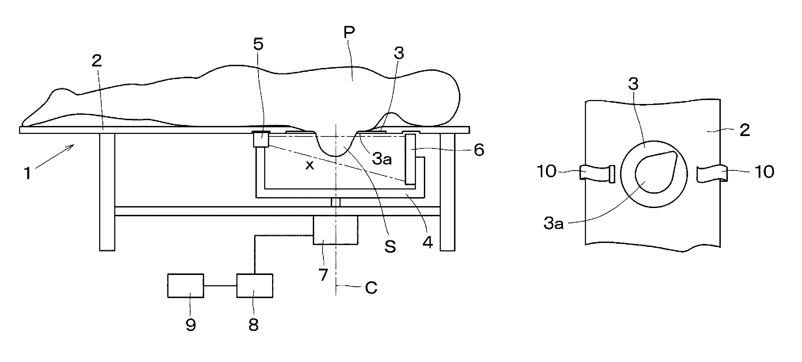
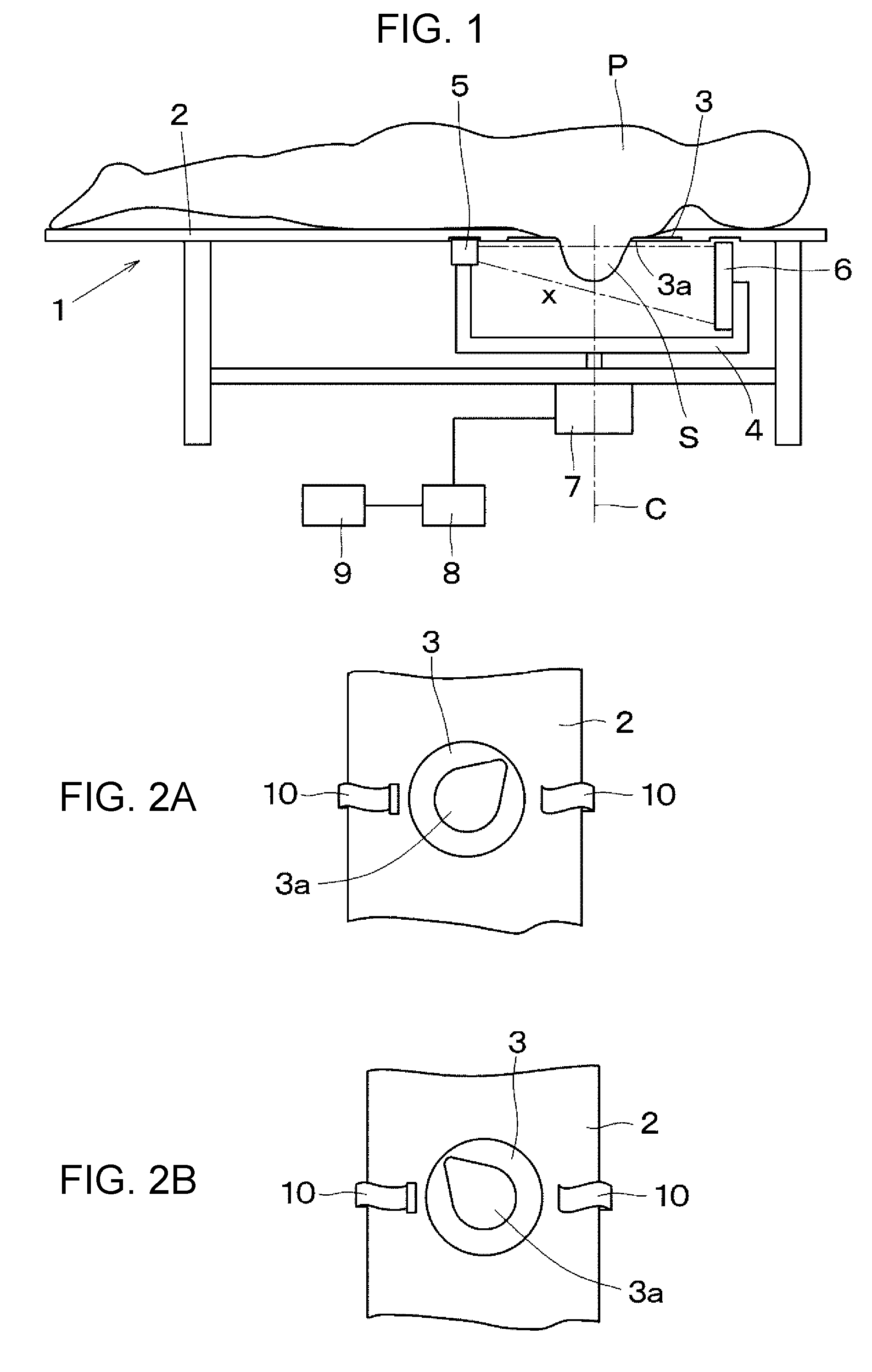
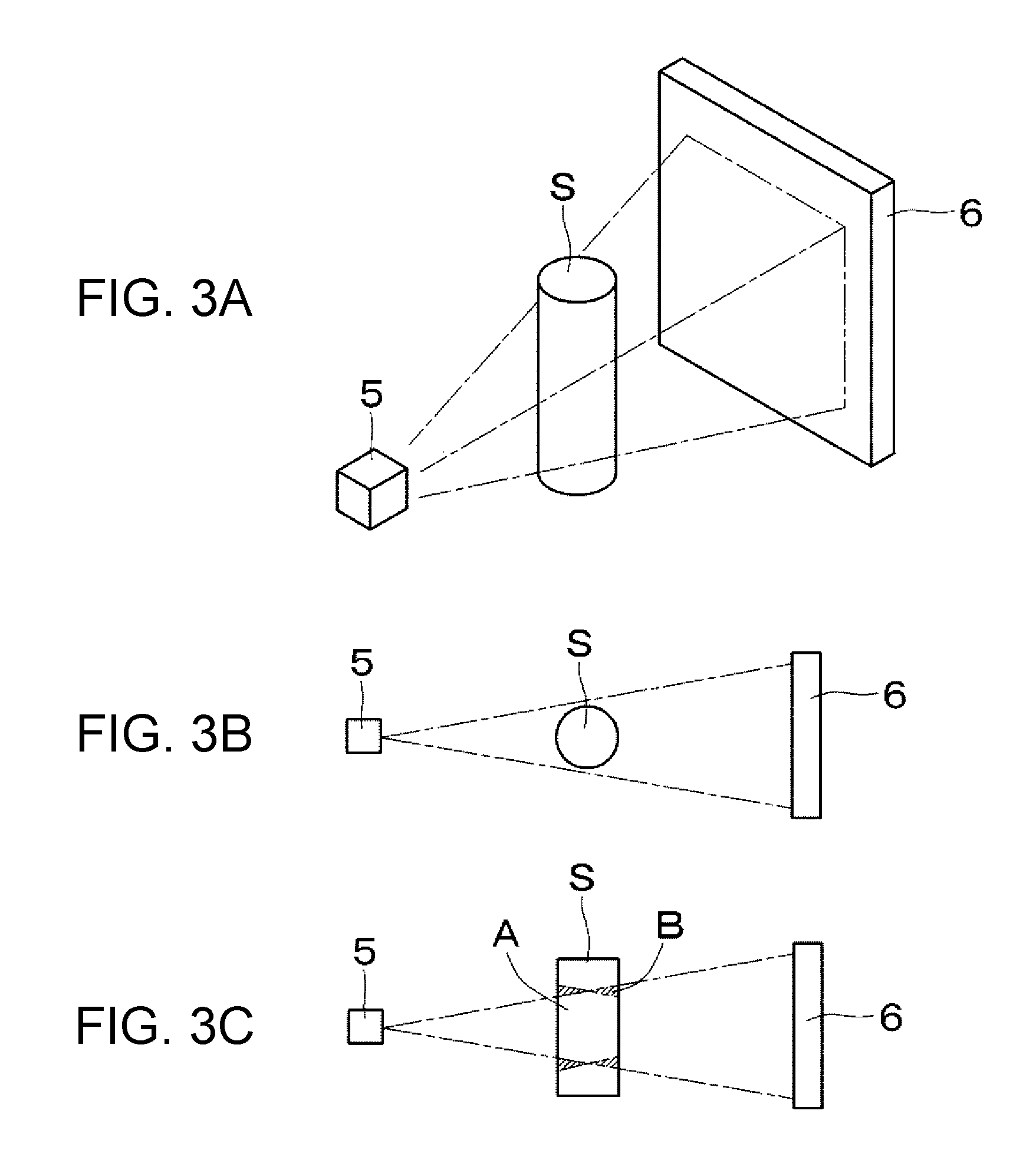
[0023]FIG. 1 shows the structure of an X-ray CT apparatus according to a first embodiment. Referring to FIG. 1, a bed 1 is used for photographing a breast of a patient P. The bed 1 includes a top plate 2 that functions as a support plate for supporting the patient P in a prone position. The top plate 2 has an aperture member 3 through which a breast of the patient P is inserted. The aperture member 3 has an opening 3a and is rotatable relative to the top plate 2 about the center of the opening 3a. The aperture member 3 is gradually tapered from the top plate 2 to the opening 3a.

[0024]An arm 4 is placed under the aperture member 3. The arm 4 rotates about a rotational axis C that extends through the center of the opening 3a in a vertical direction. An X-ray tube unit 5 and an X-ray detector 6 are respectively held at one end and the other end of the arm 4. The X-ray detector 6 includes an X-ray sensor for converting X-rays into electrical signals. The arm 4 is driven by a rotary dri...

second embodiment

[0042]FIG. 4 shows a structure of a second embodiment. Referring to FIG. 4, an X-ray tube unit 5 and an X-ray detector 6 are placed in a donut-shaped frame body 11 that is retained by a main frame (not shown). Photographing can be performed while a patient P is in an upright position by placing a breast, which is a photographing subject S, along a horizontal central axis C of the frame body 11. A central space 12 around the central axis C of the frame body 11 functions as a photographing area. The size of the frame body 11 can be reduced by limiting the photographing subject to breasts.

[0043]The X-ray tube unit 5 and the X-ray detector 6 are retained by retaining members 13 and 14, respectively, in the frame body 11. The retaining members 13 and 14 rotate about the central axis along the inner periphery of an annular guide 15. Thus, data of the breast, which is the photographing subject S, in the range of 360° can be obtained.

[0044]An aperture member 16 having an opening 16a is atta...

third embodiment

[0054]FIG. 7 is a sectional view of a frame body 11 taken along a plane perpendicular to a rotational axis according to a third embodiment. A biopsy device 22 is attached to a guide 15 together with an X-ray tube unit 5 and an X-ray detector 6 such that the biopsy device 22 can move along the guide 15. Accordingly, a needle can be inserted into the breast at an arbitrary angle to extract a biopsy sample.

[0055]In the third embodiment, X, Y, and Z coordinates of a lesion that are necessary for guiding the biopsy needle can be obtained by a single photographing process. Since the biopsy device 22 is provided in the frame body 11, a biopsy process can be performed after the X-ray photographing process without changing the state of the breast. As a result, time required for the test and burden on the patient P can be reduced.

[0056]In the above-described embodiments, X-ray is explained as an example of radiation. However, other kinds of radiations can also be used.


PUM


Abstract

A medical breast-image capturing apparatus includes an aperture member for exposing a breast of a test subject. To perform adequate breast cancer screening, the aperture member has a noncircular shape, is arranged to be replaceable with another aperture member, or has a variable opening. Another medical breast-image capturing apparatus includes a pressure-reducing device for reducing a pressure in a hollow section in which a breast is exposed.

Description

BACKGROUND OF THE INVENTION[0001]1. Field of the Invention[0002]The present invention relates to a medical breast-image capturing apparatus which acquires a medical image of a breast of a test subject using radiation such as X-rays, ultrasonic waves, MRI, etc.[0003]2. Description of the Related Art[0004]An improved X-ray mammography apparatus capable of photographing a breast of a patient (test subject) while the patient is in a prone position is discussed in, for example, Japanese Patent No. 2691073.[0005]In the mammography apparatus described in Japanese Patent No. 2691073, photographing is carried out while the patient lies on a bed in a prone position. The bed functions as a support platform and is provided with a breast-receiving aperture through which the patient inserts a breast. The breast that hangs down is exposed to X-rays while it is pressed between compression plates. In this apparatus, the patient is not required to be in an unnatural position and can relax during the ...

Claims


Application Information

Patent Timeline
Patent Type & Authority Patents(United States)
IPC IPC(8): A61B6/04
CPCA61B6/0414A61B6/502A61B6/0435
Inventor KOBAYASHI, MASAAKI
Owner CANON KK
Why Eureka
Try Eureka