
Rapid 3-dimensional bilateral breast MR imaging

a breast mr imaging and rapid technology, applied in the field of rapid, bilateral projection reconstruction methods, can solve the problems of poor image quality, affecting the diagnosis of breast cancer, and not being as accurate, and achieve the effect of rapid detection of cancer

Active Publication Date: 2012-03-13
THE TRUSTEES OF THE UNIV OF PENNSYLVANIA
View PDF6 Cites 24 Cited by
  • Summary
  • Abstract
  • Description
  • Claims
  • Application Information

AI Technical Summary

Benefits of technology

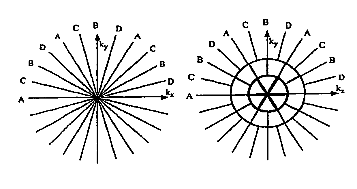
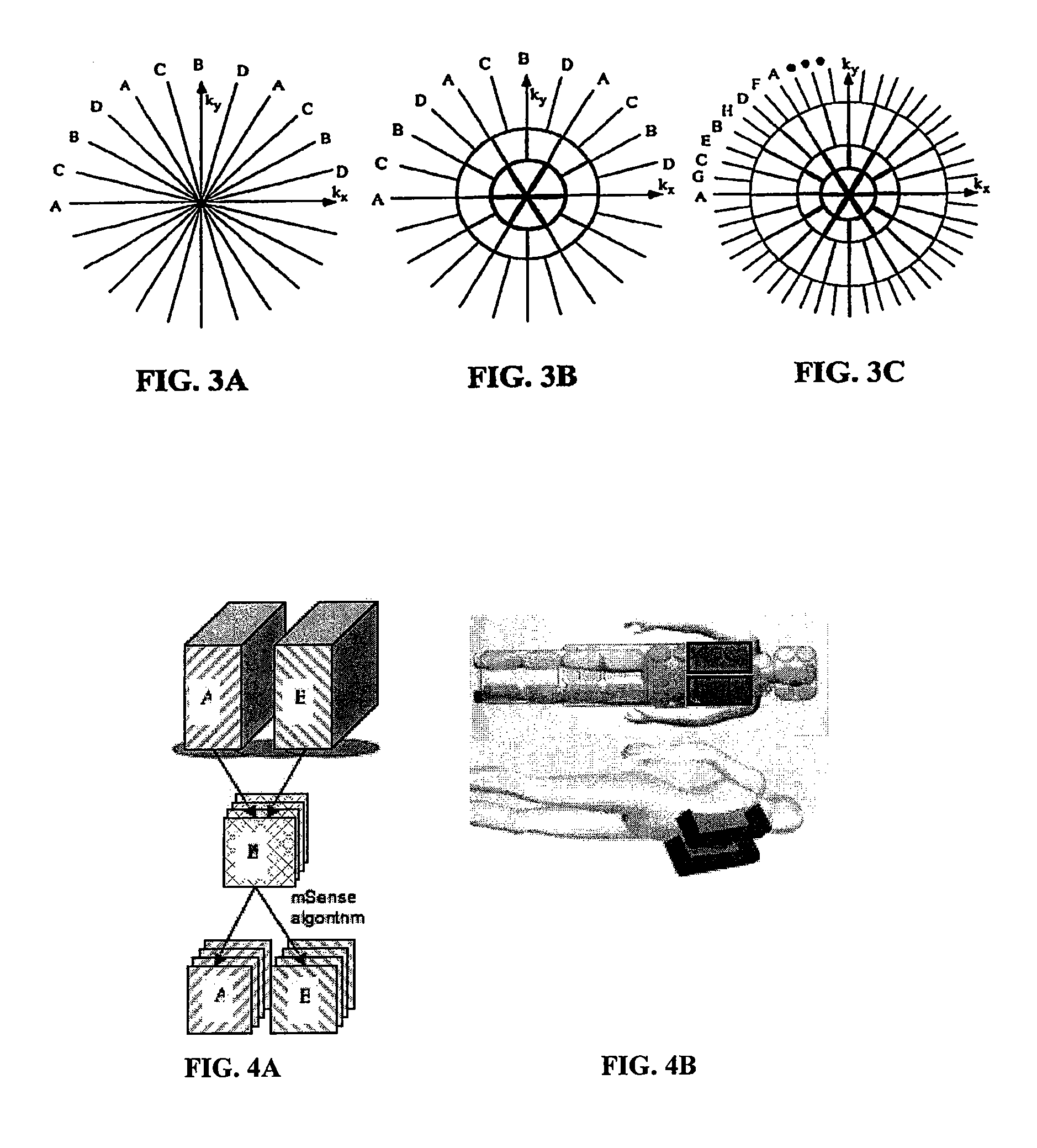
[0019]In light of the foregoing stated needs in the art, the present invention provides a bilateral projection reconstruction method for 3D dynamic contrast-enhanced MR imaging of both breasts simultaneously of an individual. “Projection reconstruction” (PR) is a method of image reconstruction that uses views of an object acquired at different angles (“radial acquisition”). Using a unique sequence approach, this new method comprises at least three elements: radial sampling, k-space weighted image contrast (KWIC), and simultaneous excitation. Thus, the present invention adds parametric mapping (color overlay) and a viewer / analysis program to evaluate the multidimensional data using the pulse sequence / reconstruction / heuristic model, thereby providing the small set of images from which radiologists were able to rapidly identify cancers in a manner not previously possible.
[0021]An initial evaluation of these methods was performed on a cohort of women presenting with palpable or mammographically visible breast abnormalities. Seventy-three abnormalities were found in 45 of the 54 bilateral examinations that were performed, and in 11 of those cases, there was a significant finding in the contralateral breast. Thus, it was confirmed that dynamic contrast enhanced images of both breasts can be acquired, simultaneously providing high-resolution images, as well as rapid sampling of the contrast kinetics.
[0022]The present high resolution bilateral MR imaging techniques offer significant advantages in the field of breast cancer screening, including, e.g.: (1) the improved ability to detect early breast cancers; (2) the improved ability to differentiate between breast cancer and normal breast tissue; (3) the improved ability to differentiate between breast cancer and benign lesions in the breast; (4) the improved ability to differentiate between scar and recurrent breast cancer; (5) the improved detection of multifocal and / or multicentric breast cancers; (6) the improved ability to determine the extent of breast cancer present; (7) the improved ability to detect early breast cancer in individuals with breast implants; (8) the improved ability to detect breast cancer in individuals with breast implants while also evaluating the breast implants for abnormalities; and (9) the improved ability to detect early breast cancer in individuals with dense breasts.

Problems solved by technology

Such misses have been attributed to the subtle nature of the visual findings, poor image quality, or oversight by the radiologist.
Mammography is also disadvantageous in that it is limited in individuals who have breast implants, and is not as accurate in younger individuals whose breast tissue tends to be denser.
In addition, mammography exposes individuals to ionizing radiation that may increase their risk of developing breast cancer.
Mammography also requires significant compression of the breast tissue that many individuals find painful, leading them to avoid mammography.
However, the smaller the size of the local coil, the smaller its field of view, or sensitivity profile.
However, one of the challenges associated with using multiple coils for imaging is the fact that the fields of individual coils may interact, resulting in coil-to-coil coupling.
Such interactions tend to reduce the coil quality factor, or Q.
However, even for a unilateral breast study, the simultaneous acquisition of both high spatial resolution data for architectural analysis and high temporal resolution data for contrast kinetic classification is difficult due to their diverging demands (Dougherty et al., ISMRM 13th Scientific Meeting and Exhibition, page 86, Miami Beach, May 2005).
The high-resolution imaging needed to distinguish features necessary for architectural interpretation requires a relatively long time to acquire.
Acquiring fewer slices or reducing the matrix size will speed acquisition but at the cost of coverage or spatial resolution.
However, this acquisition scheme causes the mixing of the constantly updated low spatial frequency data with the high frequency data acquired at different time periods, potentially resulting in blurring of the enhancing structures in the phase encoding direction.
However, the inevitable mixing of old and new data that are non-contiguous in time still occur, and may cause measurement errors.
(J. Magn. Reson. Imaging 13:313-317 (2001A)) have previously described the use of multi-coil arrays for separation of signals from multiple, simultaneously excited slices, but was not adapted to multiple 3D volumes.
However, with greater acceleration factors the SNR is concomitantly decreased and the time resolution is still insufficient to adequately sample the contrast kinetics.
However, the main drawback of this technique is also its strength—that is, the temporal / spatial resolution tradeoff.
Due to SNR limitations and the desire for artifact-free images, high spatial resolution images required lower temporal resolution since a greater number of views were needed during reconstruction.
However, the problem of imaging at a high frame rate while preserving spatial resolution is compounded in the case of bilateral imaging.
In a clinical bilateral exam the acquisitions are often interleaved, which doubles the scan repetition rate (TR) and reduces the temporal resolution.
In another approach often used clinically, the breasts are scanned with individual unilateral studies on consecutive days with a significant penalty in cost and patient inconvenience.
As a result, while these methods offered a significant improvement, alone they cannot achieve the temporal resolution needed to adequately sample the enhancement curve, while simultaneously acquiring high spatial resolution images of both breasts.

Method used


Image

  • Rapid 3-dimensional bilateral breast MR imaging
  • Rapid 3-dimensional bilateral breast MR imaging
  • Rapid 3-dimensional bilateral breast MR imaging

Examples


example 1

[0101]Using the methods described herein, fifty-four (54) bilateral exams were performed on women with breast abnormalities, all with excellent image quality. For each case, 64 high-resolution slices (0.47×0.47 mm / pixel) and 25 temporal phases (5 baseline and 20 post-contrast) were reconstructed. The scan repetition time was set at 9.8 ms in this study in order to be compatible with a previous breast imaging study. The minimum TR for the sequence, at the same bandwidth, was actually 8.1 milliseconds, which provided an effective temporal resolution of 12.4 seconds. Given sufficient SNR, from, e.g., a high field scanner, the receiver bandwidth could be increased and the TR reduced even further to improve the temporal resolution. All reconstruction, image analysis and viewing were performed on a dual Xeon processor PC at 3.1 GHz.

[0102]Total time for each volunteer to be in the scanner was ˜25 minutes, including patient set-up. FIGS. 6A-6B show a representative case including post-contr...

example 2

[0108]In light of the foregoing findings that rapid radial dynamic contrast enhanced (DCE) MR breast imaging provides high spatial resolution for architectural assessment, as well as high temporal resolution for better characterization of the kinetic response, lesion characterization was correlated with histopathologic findings to determine the diagnostic performance. The kinetic assessment of breast tumors using the signal enhancement ratio (SER) (Li et al., Magn. Reson. Med, 58:572-581 (2007)) has been shown to be a predictor of malignant disease, while being independent of the T1 relaxation time and image intensity scaling. Accordingly, in the present example, DCE-MR images of the breasts were acquired using an undersampled radial trajectory and the kinetic response was assessed using SER.

[0109]Methods: One hundred twenty six (126) subjects with palpable or mammographically-visible suspicious findings were recruited and granted IRB approval. From this number, 94 had subsequent pa...


PUM


Abstract

Provided is a method for rapid, 3D, dynamic, projection reconstruction bilateral breast imaging using simultaneous multi-slab volume excitation and radial acquisition of a contrast enhanced bilateral image, in conjunction with SENSE processing, using k-Space Weighted Image Contrast (“KWIC”) filtering and multi-coil arrays for signal separation in an interleaved bilateral MR bilateral breast scan that uses conventional Cartesian sampling without parallel imaging. Software was developed for the reconstruction, modeling contrast kinetics using a heuristic model, display by parametric mapping and viewer / analysis of the multidimensional, high frame-rate bilateral breast images.

Description

CROSS REFERENCE TO RELATED APPLICATIONS[0001]This application is a continuation of International Application PCT / US2007 / 009824 filed Apr. 23, 2007, published as WO 2007 / 124151, which claims priority to U.S. Provisional Application No. 60 / 793,799 filed Apr. 21, 2006, each of which is incorporated herein in its entirety.GOVERNMENT INTEREST[0002]This invention was supported in part by Grant No. R01-CA-90699 from the National Institutes of Health. Accordingly, the Government may have certain rights in this invention.FIELD OF THE INVENTION[0003]The present invention relates to a rapid, bilateral projection reconstruction method for 3D dynamic contrast-enhanced Magnetic Resonance Imaging (“MRI” or “MR imaging”) of both breasts of an individual simultaneously, providing high-resolution images, as well as rapid sampling of the contrast kinetics.BACKGROUND OF THE INVENTION[0004]Breast cancer is the most frequently diagnosed malignancy among women other than skin cancer, and the second leadin...

Claims


Application Information

Patent Timeline
Patent Type & Authority Patents(United States)
IPC IPC(8): A61B5/05
CPCA61B5/055A61B5/415A61B5/418G01R33/4824G01R33/5601G01R33/5611G01R33/4828G01R33/56308
Inventor DOUGHERTY, LAWRENCESONG, HEE KWONBOSTON, RAYMOND CHARLESSCHNALL, MITCHELL
Owner THE TRUSTEES OF THE UNIV OF PENNSYLVANIA
Learn More