
Rigid arthroscope system

a rigid, arthroscope technology, applied in the field of arthroscopic surgical instruments, can solve the problems of shortening the useful life of the prosthetic, rigid cannula can become stuck between bones or cartilage, and scratching or gouge soft tissue within the operating field of the rigid cannula, so as to achieve the effect of enduring high torque and bending stress and being sufficiently rigid

Active Publication Date: 2012-07-24
PRISTINE SURGICAL LLC
View PDF21 Cites 18 Cited by
  • Summary
  • Abstract
  • Description
  • Claims
  • Application Information

AI Technical Summary

Benefits of technology

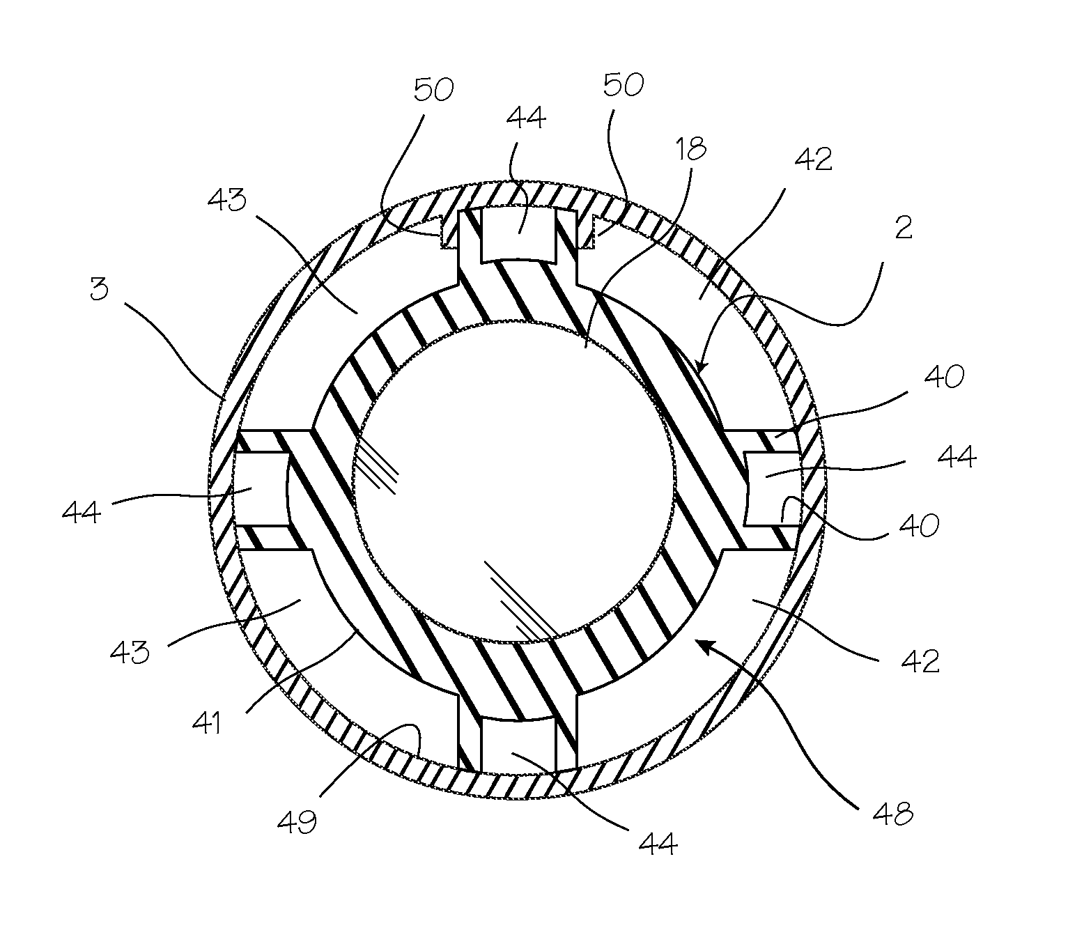
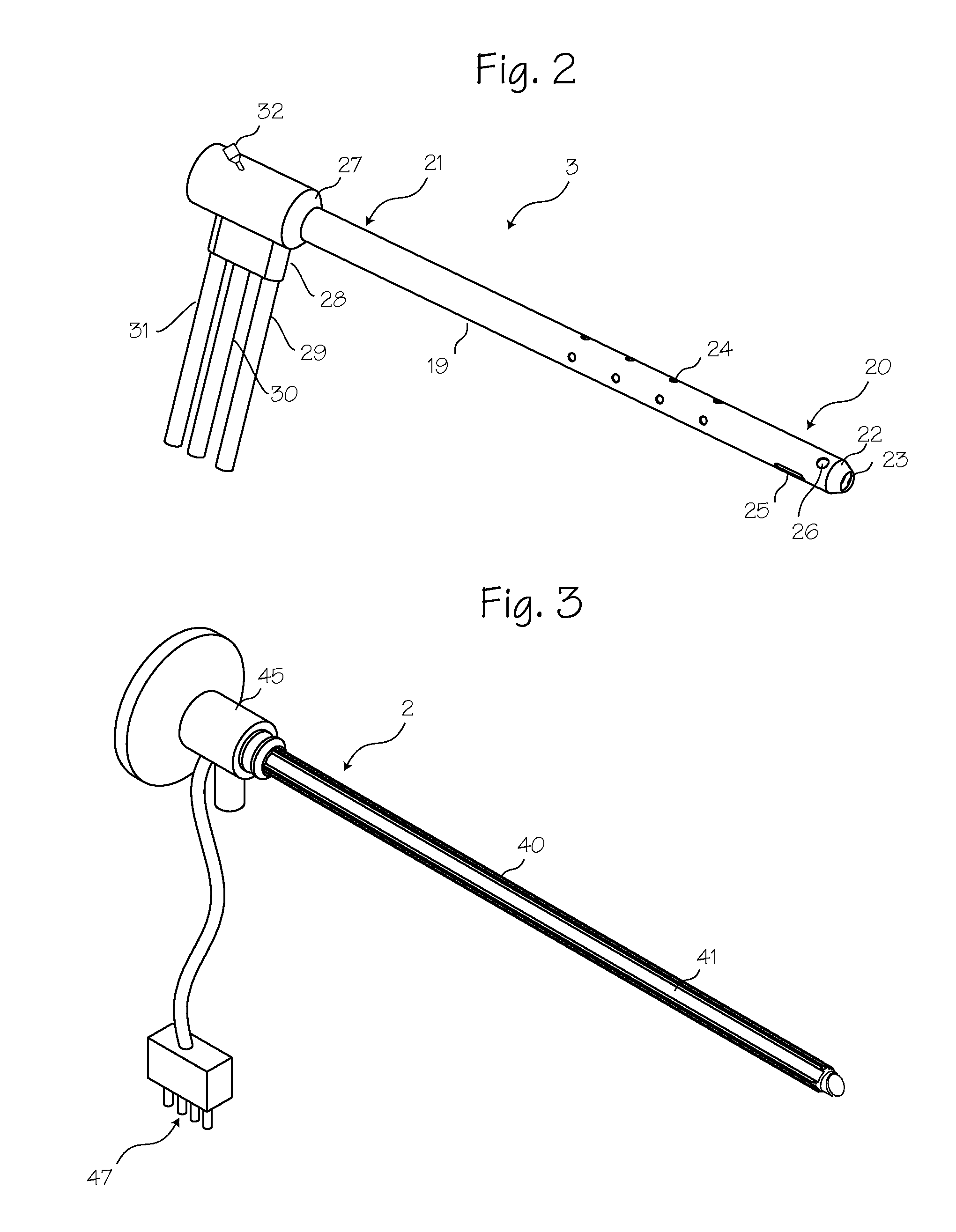
"The arthroscope described in this patent is a reinforced endoscope that has separate channels for fluid management. It is designed to be rigid enough to penetrate and move within a joint while withstanding high levels of torque and bending stress. The external sheath is made of a simple polymer sheath that protects the joint tissue from unnecessary instrument trauma and scuffing. The sheath is disposable and helps clean and sterilize the scope between uses. The arthroscope architecture provides the surgeon with the feel of a traditional metal instrument and multiple fluid management channels in a low-profile, small diameter system. The arthroscope may also have permanently mounted sensors such as temperature and pressure sensors."

Problems solved by technology

The distal end of the rigid cannula is pointed, usually sharp, and thus the rigid cannula can scratch or gouge soft tissue within the operating field.
The rigid cannula can also become stuck between bones or cartilage during a procedure.
The metal surface of the rigid cannula can also damage metal prosthetics used to replace joints, resulting in a shortening of the useful life of the prosthetic and forcing the patient to undergo additional, painful surgeries to correct the problem.
An additional problem associated with arthroscopic surgery is maintaining a clear surgical field during surgery.
Blood and debris can cloud the field, impairing a surgeon's ability to visualize tissue.
Excessive force on the sheathed endoscope may crush or shear off the plastic ribs on the inner side of the sheath, thus destroying the fluid channels.
Excessive force can also break the scope, requiring very expensive repairs or replacement of the scope.

Method used


Image

  • Rigid arthroscope system
  • Rigid arthroscope system
  • Rigid arthroscope system

Examples


Embodiment Construction

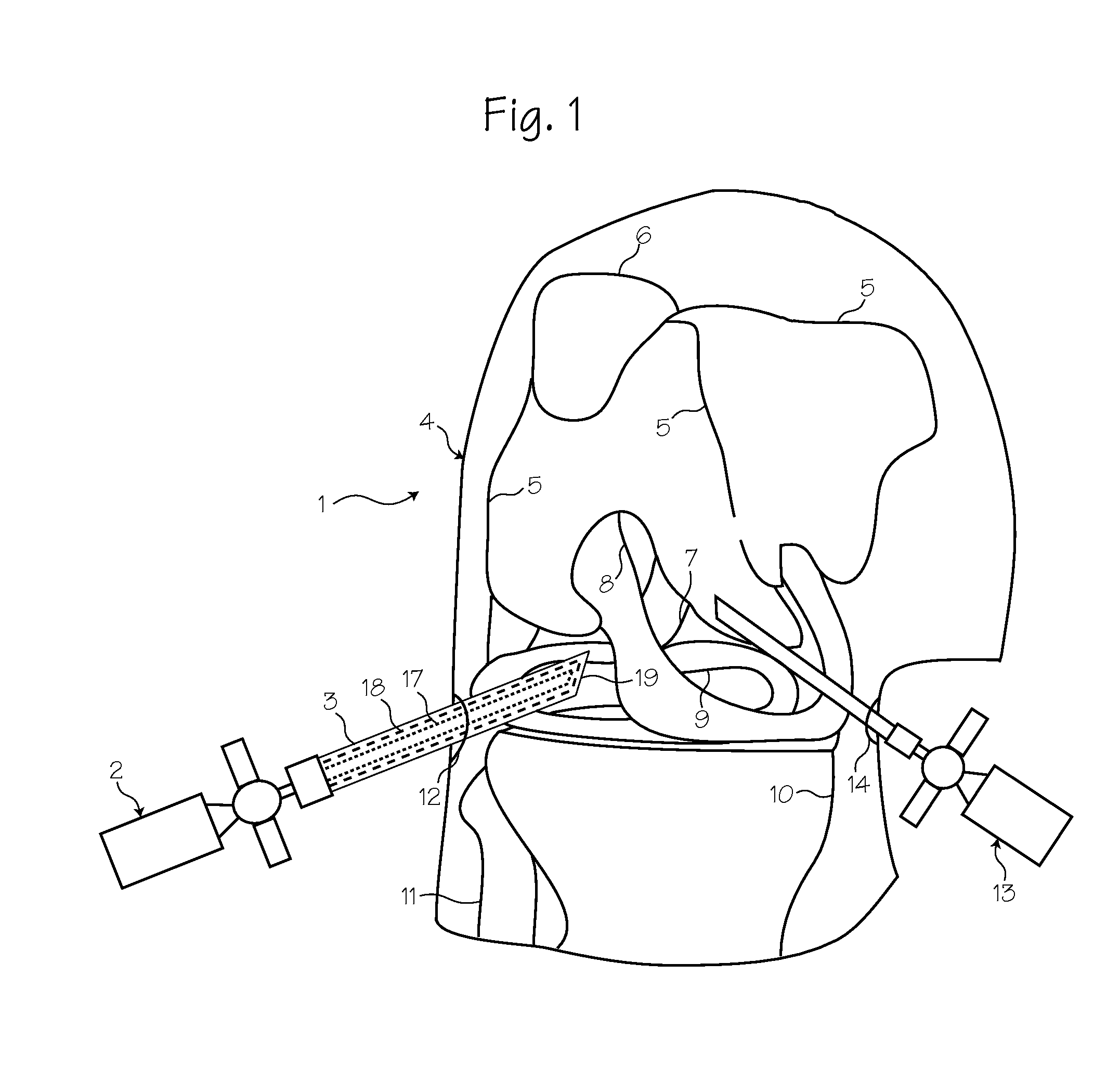
[0017]FIG. 1 shows a method of performing arthroscopic surgery on a patient 1 using an arthroscope 2 sheathed in an atraumatic sheath 3. In FIG. 1, the various parts of the arthroscope are shown in phantom to indicate their positions inside the sheath. Various anatomical landmarks in the patient's knee 4 are shown for reference, including the femur 5, patella 6, posterior cruciate ligament 7, anterior cruciate ligament 8, meniscus 9, tibia 10 and fibula 11. During surgery, the surgeon introduces the arthroscope 2 into the knee via a first incision 12 in order to visualize the surgical field. A trimming instrument 13 is introduced through a second incision 14 to remove or trim tissue that the surgeon determines should be removed or trimmed.

[0018]The arthroscope 2 is an optical instrument comprising an optical rod 17 surrounded by a rigid tube 18. To protect the patient from unintended injury or trauma during the procedure, the arthroscope has been inserted into a resilient, outer int...


PUM


Abstract

A reinforced arthroscope comprising external ribs to provide for a number of separate fluid channels, such as inflow, outflow and interstitial tissue drainage, when the arthroscope is slipped into a disposable external sheath. The arthroscope is constructed to be sufficiently rigid so as to penetrate and move within a joint without damaging the rod optics inside. The externality of the arthroscope channels allow for cleaning and sterilizing the scope between uses.

Description

FIELD OF THE INVENTIONS[0001]The inventions described below relate to the field of arthroscopic surgical instruments.BACKGROUND OF THE INVENTIONS[0002]Arthroscopic surgery involves using optical instruments, such as an arthroscope, to visualize an operating field inside or near a joint of a patient. The same instrument or other instruments may be used to perform a surgical procedure in the operating field. Common instruments used in addition to the arthroscope include a trimming instrument for cutting tissue and an irrigation instrument for irrigating the surgical field. Each of the instruments requires its own incision to be introduced into the surgical field. Thus, many surgeons prefer to use only a trimming instrument and an arthroscope during arthroscopic surgical procedures.[0003]Arthroscopes are fragile in relation to the forces applied during arthroscopic surgery, so a rigid cannula is placed over the arthroscope to reinforce it. The distal end of the rigid cannula is pointed...

Claims


Application Information

Patent Timeline
Patent Type & Authority Patents(United States)
IPC IPC(8): A61B1/012
CPCA61B1/317A61B1/015A61B1/00078A61B1/00135A61B17/3417A61B17/3421A61B2017/00092A61B2217/007A61B17/3474A61B2019/465A61B2090/065A61B1/00073A61B1/018A61B1/126
Inventor KUCKLICK, THEODORE R.
Owner PRISTINE SURGICAL LLC
Learn More