
Cervix monitoring system and related devices and methods

Inactive Publication Date: 2005-03-03
MANSOUR HEBAH NOSHY +1
View PDF24 Cites 141 Cited by
  • Summary
  • Abstract
  • Description
  • Claims
  • Application Information

AI Technical Summary

Benefits of technology

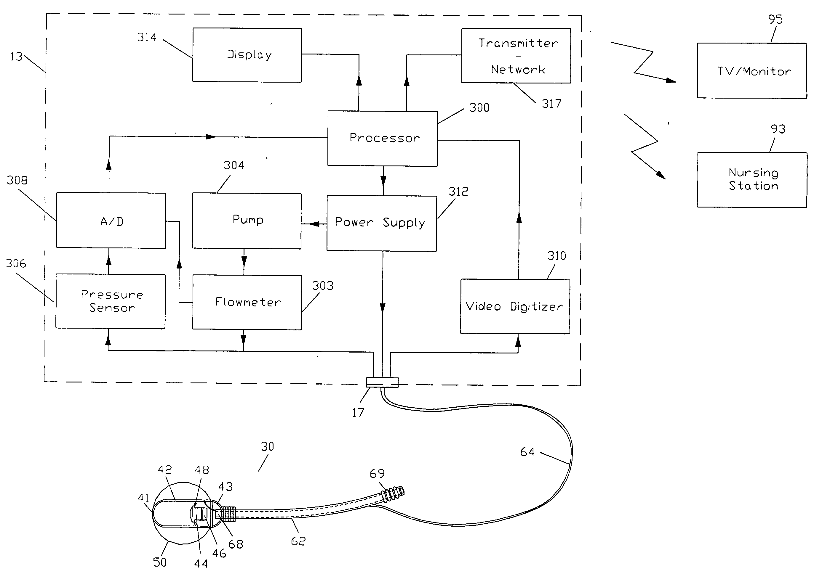
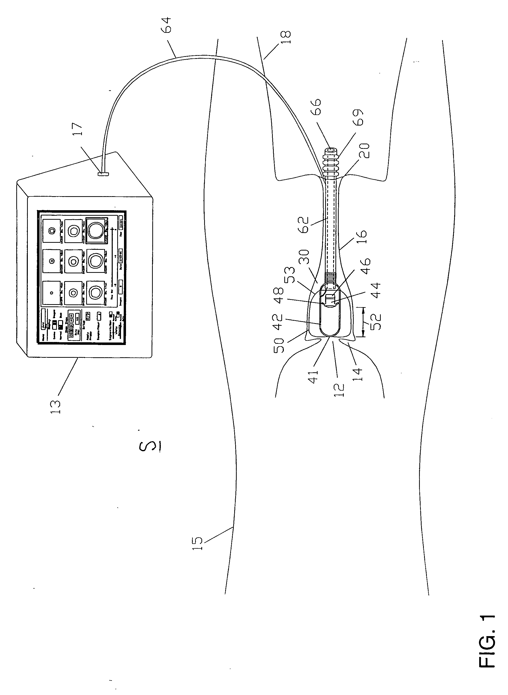
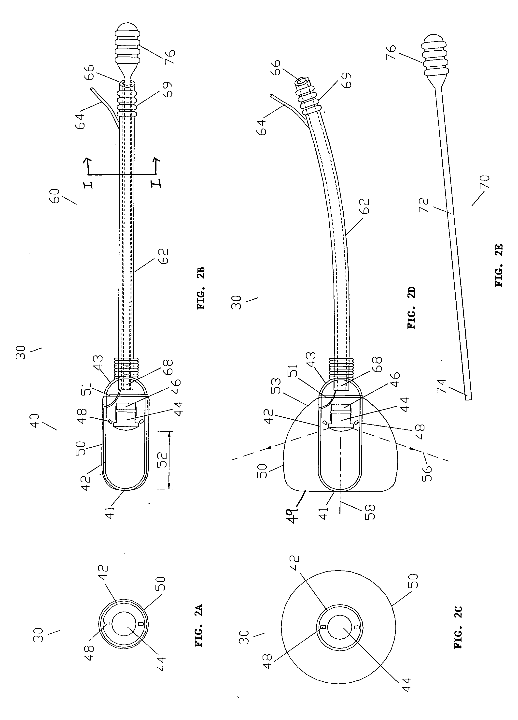
The present invention is directed to a minimally invasive system and method for monitoring changes in a cervix opening during labor, including changes in its diameter. The system having a probe and a monitoring unit serve to monitor periodically the cervix opening during labor and obtain measurements of the diameter of the cervix opening. The probe primarily includes a camera for imaging the cervix opening, a lens to provide an optimal field of view for the camera at close range to the cervix opening, a light source to illuminate the cervix and a balloon to expand the vagina around the probe and position the probe to allow unobstructed imaging of the cervix. The balloon is triggered to inflate when images of the cervix opening are taken and can deflate when the camera is not capturing images. A tail or handle portion of the probe facilitates the insertion and removal of the probe from the vagina and protects electrical and pneumatic connections between the probe and the monitoring unit. The tail portion is generally flexible but is adapted to have sufficient rigidity by means of a rod applicator, bead trains or an inflatable body to facilitate the insertion of the probe into the vagina.
In one embodiment, the processor implements segmentation techniques and / or blob analysis algorithms to analyze the images of the cervix to identify the opening and measure its diameter. Other algorithms may also be implemented to correct for any spatial distortion, particularly barrel distortion. In another embodiment, a portable controller is provided to facilitate the mobility of the patient, by eliminating a cable extending between the probe and the monitoring unit.

Problems solved by technology

This method is known as digital probing and suffers several inherent limitations, including the following.
Second, the method can cause discomfort to the patient during each session of digital probing which may be performed repeatedly at an interval of 30 minutes or so throughout labor.
Third, the health care provider may forget to perform the exam or even avoid it intentionally if the patient is sleeping.
Fourth, hand examinations can increase the risk of infection to the patient and the fetus especially if the fetal membranes have been ruptured.
In the latter regard, hand examinations may cause complications, including neonatal sepsis to the infant, chorioamnionitis to the fetal membranes, and / or endomyometritis to the uterine muscles.
As such, hand examinations are often minimized and hence do not provide continuous reliable monitoring of the progress of the labor.
(Lucidi, R. Scott; Blumenfeld, Lee A.; Chez, Ronald A., “Cervimetry: A Review of Methods for Measuring Cervical Dilatation During Labor,” Obstetrical and Gynecological Survey, Volume 55(5), pp 312-320, May 2000) who wrote “although many instruments have been developed to measure cervical dilatation during labor and their use as a research tool has been established, no device has yet been successfully used for clinical obstetrics.
The ideal device has not yet been developed; however, because repeated digital cervical examinations are time consuming for the clinician, are poorly reproducible, and are uncomfortable for the patient, continued efforts to develop a cervimeter suitable for clinical use is a worthwhile endeavor.”
However, previous techniques failed to gain clinical acceptance due to several limitations, including the complexity of installation, inaccuracy of measurements, tissue trauma caused by the devices or their components, including the manner by which the components are attached to the cervix, blockade of the cervical canal, costly sterilization between uses, and / or patient discomfort.

Method used


Image

  • Cervix monitoring system and related devices and methods
  • Cervix monitoring system and related devices and methods
  • Cervix monitoring system and related devices and methods

Examples


Embodiment Construction

Referring to FIGS. 1 and 6, the present invention includes a system S for monitoring dilation of an opening 12 of a cervix 14 of a female patient 15 who is pregnant and in labor to deliver. The system includes a probe 30 that is inserted into a vagina 16 of the patient to gather data relating to the cervix, in particular, the diameter of the opening, and a monitoring unit 13 that is in communication with the probe and receives data from the probe which is processed by a processor to provide and display useful information to health care providers located proximately to the patient and / or remotely therefrom. Advantageously, the probe 30 is adapted to remain in the patient's vagina throughout the duration of labor and assume a suitable position in the vagina so as to gather image data that is sent to the monitoring unit. Accordingly, the patient need not have her cervix digitally probed during labor and may remain relatively undisturbed while changes in the size of the cervix opening ...


PUM


Abstract

The present invention is directed to a minimally invasive system and method for monitoring changes in a cervix opening during labor, including changes in its diameter. The system having a probe and a monitoring unit serve to monitor periodically the cervix opening during labor and obtain measurements of the diameter of the cervix opening. The probe primarily includes a camera for imaging the cervix opening, a lens to provide an optimal field of view for the camera at close range to the cervix opening, a light source to illuminate the cervix and a balloon to expand the vagina around the probe and position the probe to allow unobstructed imaging of the cervix. The monitoring unit which controls the probe to acquire images of the cervix and receives and processes image data from the probe includes a processor which uses image segmentation techniques to identify and measure the opening of the cervix and generates data signals. The monitoring unit also includes an image display which displays information based on the data signals for viewing by health care providers and / or the patient, including graphic images of the cervix opening and other text information relating to the cervix opening as an indicator of the progress of labor. In one embodiment, the processor implements segmentation techniques and / or blob analysis algorithms to analyze the images of the cervix to identify the opening and measure its diameter. Other algorithms may also be implemented to correct for any spatial distortion, particularly barrel distortion.

Description

FIELD OF THE INVENTION The present invention relates to a method and system for monitoring progress of labor during childbirth, in particular, changes in the cervix, including changes in the size of the opening of the cervix, during childbirth. BACKGROUND OF THE INVENTION Monitoring the progress of cervix dilation is desirable during labor because the progress of cervix dilation can be an indicator of various conditions or factors including: (1) arrested or dysfunctional labor, (2) cephalopelvic disproportion, (3) when and whether a Caesarean-section should be performed, (4) when and whether labor inducing / enhancing drugs (e.g. Pitocin) should be used, (5) when should different anesthetic or analgesic agents be administered, and / or (6) when the patient should begin pushing. In current clinical practice, cervix dilation (or dilatation, used interchangeably herein) is typically, if not always, monitored manually by a health care provider who inserts a gloved hand into the patient's...

Claims


Application Information

Patent Timeline
IPC IPC(8): A61B5/107
CPCA61B5/1076A61B5/412A61B5/7445A61B5/7435A61B5/435A61B5/6853
Inventor MANSOUR, HEBAH NOSHYSHEHADA, RAMEZ EMILE NECOLA
Owner MANSOUR HEBAH NOSHY
Learn More