
Therapeutic agents for diseases associated with apoptotic degeneration in ocular tissue cells that use SIV-PEDF vectors

a technology of ocular tissue cells and therapeutic agents, which is applied in the direction of biocide, drug composition, peptide/protein ingredients, etc., can solve the problems that administration methods that are expected to provide only transient effects cannot be considered suitable therapeutic methods for glaucoma, and the molecular weight of neurotrophic factors is large, so as to achieve the effect of delivering ped

Inactive Publication Date: 2012-10-02
DNAVEC CORP +1
View PDF6 Cites 5 Cited by
  • Summary
  • Abstract
  • Description
  • Claims
  • Application Information

AI Technical Summary

Benefits of technology

The present invention is about creating pharmaceutical agents to treat diseases associated with apoptotic degeneration in ocular tissue cells, such as glaucoma. The invention involves using a retroviral vector, specifically a simian immunodeficiency virus vector carrying a pigment epithelium derived factor gene, to effectively deliver the therapeutic gene to the retina. The vector is designed to be safe and effective, with the ability to protect ganglion cells and reduce side-effects. The invention also includes a method for administering the pharmaceutical agent and a method for producing the vector. Overall, the invention provides a solution for delivering therapeutic agents to treat glaucoma and other ophthalmic diseases associated with apoptotic degeneration.

Problems solved by technology

However, glaucoma can lead to blindness if not appropriately treated.
It is difficult to continuously deliver large molecular weight proteins to the retina using the current drug delivery systems.
Considering that glaucoma is a chronic disease, administration methods that are expected to provide only transient effects cannot be considered as suitable therapeutic methods for glaucoma.
On the other hand, retroviral vectors may generally enable long-term expression of genes by being stably incorporated into the chromosomes of dividing cells; however, there is so far no known study of glaucoma therapy that uses retroviral vectors into which PEDF has been inserted.[Patent Document 1] International Application No.

Method used


Image

  • Therapeutic agents for diseases associated with apoptotic degeneration in ocular tissue cells that use SIV-PEDF vectors
  • Therapeutic agents for diseases associated with apoptotic degeneration in ocular tissue cells that use SIV-PEDF vectors
  • Therapeutic agents for diseases associated with apoptotic degeneration in ocular tissue cells that use SIV-PEDF vectors

Examples


example 1

Construction of VSV-G Pseudotyped SIV Vectors

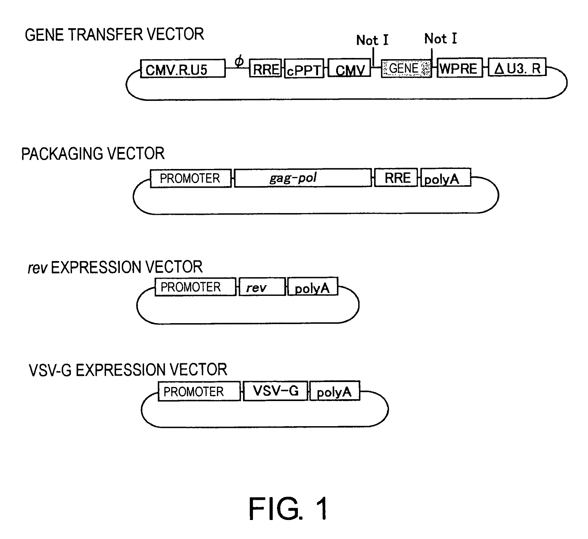
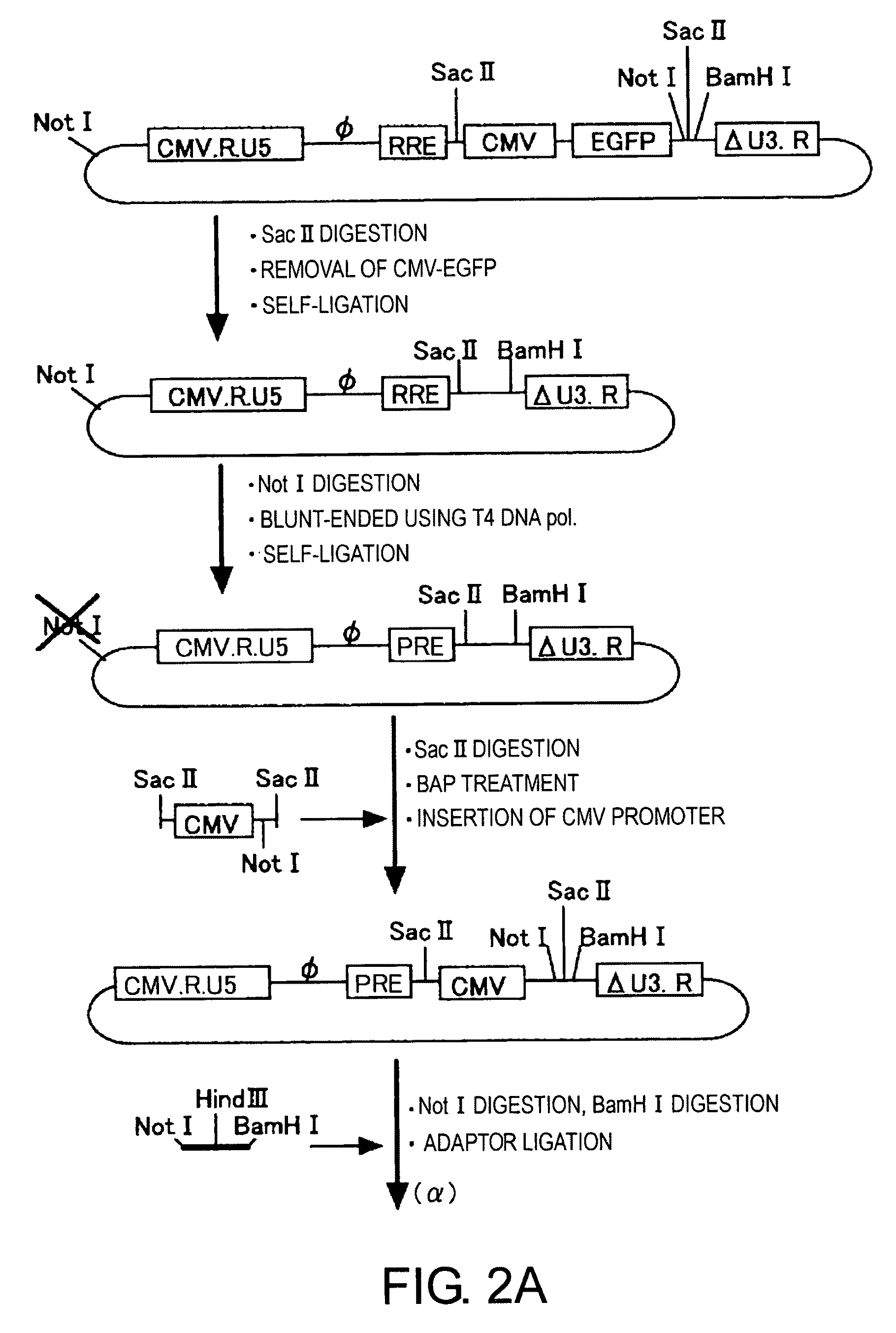
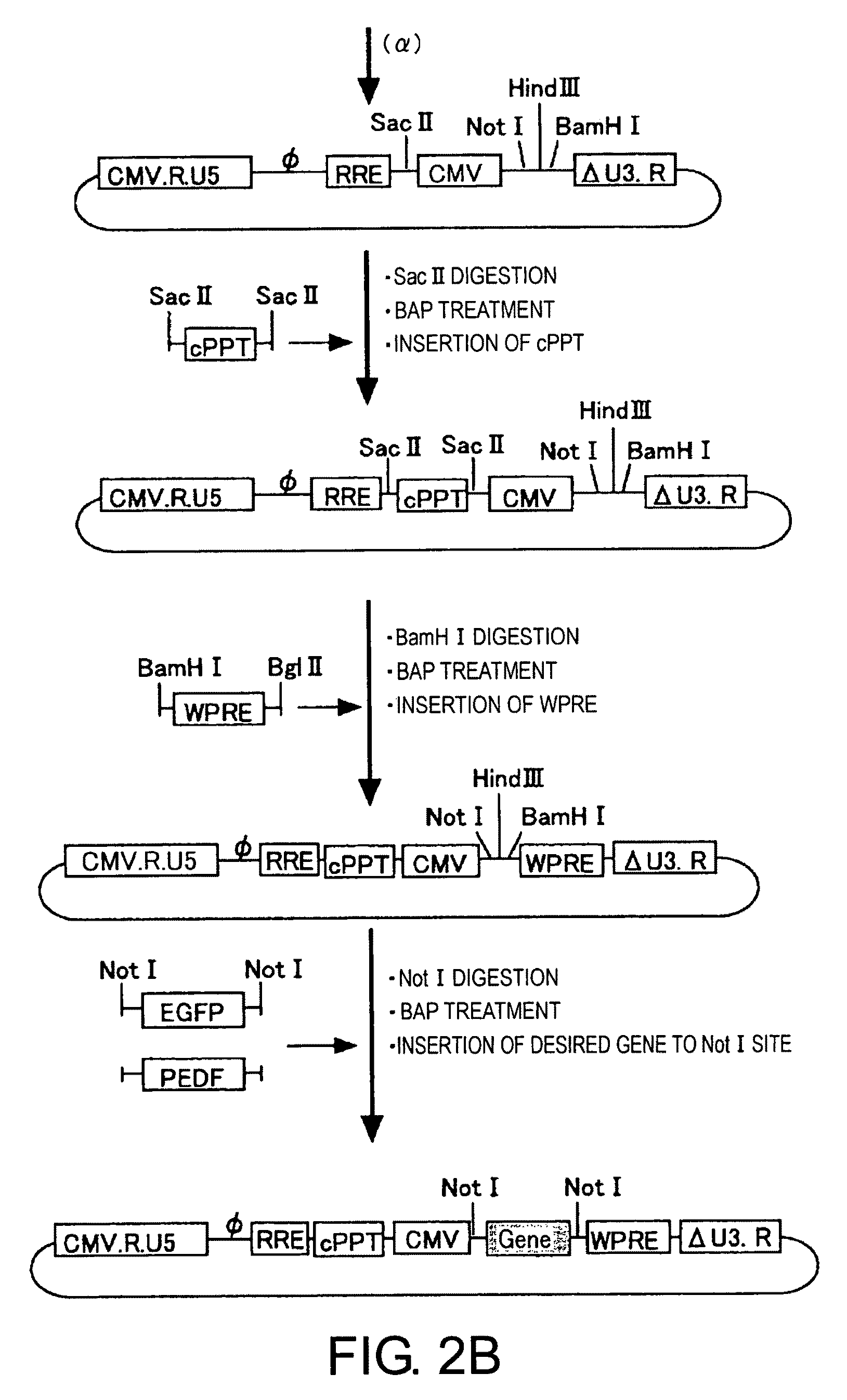
[0068]The four types of plasmids (gene transfer vector, packaging vector, rev expression vector, and VSV-G expression vector) used for vector construction are shown in FIG. 1. Three of these vectors—the gene transfer vector, packaging vector, and rev expression vector—were produced by improving conventional vector plasmids (PCT / JP00 / 03955). For the VSV-G expression vector, a conventional vector was used without modification.

[0069]Various commercially available kits were used for plasmid production. The restriction enzymes used were from New England Biolabs, and kits from QIAGEN (QIAquick PCR purification kit, QIAquick Nucleotide Removal kit, QIAquick Gel extraction kit, Plasmid Maxi kit) were used to extract, purify and recover plasmid DNAs. EX Taq enzyme from TaKaRa was used for PCR, and the primers used were synthesized by an outside manufacturer (Sigma Genosys Japan). Alkaline phosphatase (E. coli C75) from TaKaRa was used for dephosph...

example 2

Evaluation of Function of the SIV Vector Carrying cPPT and WPRE

[0084]To investigate the effect of the introduced cPPT and WPRE, vectors carrying cPPT or WPRE alone were produced as well as those carrying cPPT and WPRE simultaneously, and these were compared to the conventional type control. All gene transfer vectors used carried EGFP. The packaging vector used was a conventional type (SEQ ID NO: 27).

2-1. Preparation of SIV Vectors

[0085]Human fetal kidney cell-derived cell line 293T cells were plated in 15-cm plastic dishes at approximately 1×10⁷ cells per dish (a density to reach 70-80% on the following day) and cultured for 24 hours in 20 mL of D-MEM medium (Gibco BRL) containing 10% fetal calf serum. After culturing the cells for 24 hours, the medium was replaced with 10 mL of OPTI-MEM medium (Gibco BRL), and the cells were used for transfection.

[0086]After 6 μg of the gene transfer vector, 3 μg of the packaging vector, and 1 μg of the VSV-G expression vector were dissolved in 1.5...

example 3

Large-Scale Preparation and Concentration of SIV Vectors Carrying Therapeutic Genes

[0095]An SIV vector was produced as described below based on four types of plasmids shown in FIG. 1: the improved gene transfer vector, packaging vector, rev expression vector, and VSV-G expression vector. The vector carrying the therapeutic gene PEDF was produced in a set of twenty 15-cm dishes.

[0096]293T cells were plated in 15-cm plastic dishes at approximately 1×10⁷ cells per dish (a density to reach 70-80% on the following day) and cultured for 24 hours in 20 mL of D-MEM medium containing 10% fetal calf serum. After culturing the cells for 24 hours, the medium was replaced with 10 mL of OPTI-MEM medium, and the cells were used for transfection. After dissolving 10 μg of a gene transfer vector, 5 μg of packaging vector, 2 μg of rev expression vector, and 2 μg of VSV-G expression vector in 1.5 mL of OPTI-MEM medium per dish, 40 μL of PLUS Reagent (Invitrogen) was added and stirred. Then the mixture...


PUM

PropertyMeasurementUnit
intraocular pressureaaaaaaaaaa

Abstract

The present invention provides novel methods for treating diseases associated with apoptotic degeneration in ocular tissue cells by effective administration of pigment epithelium derived factor (PEDF). The present inventors studied PEDF as a means to prevent ganglion cell death, the final pathology of glaucoma. The present invention is particularly focused on SIV vectors for effective methods for delivering PEDF, and constructed an SIV-PEDF vector. When the SIV-PEDF vector was administered subretinally to an ischemia reperfusion model and NMDA-induced model, a significant suppression effect on ganglion cell death was observed. The present inventors therefore discovered that the SIV-PEDF vector is an effective pharmaceutical agent for treating diseases associated with apoptotic degeneration in ocular tissue cells, such as glaucoma.

Description

CROSS-REFERENCE TO RELATED APPLICATIONS[0001]This application is the U.S. National Stage of International Application PCT / JP2006 / 303032, filed Feb. 21, 2006, which claims the benefit of Japanese Application Serial No. JP 2005-047951, filed Feb. 23, 2005.TECHNICAL FIELD[0002]The present invention relates to methods for treating glaucoma using a lentiviral vector carrying a neurotrophic factor.BACKGROUND ART[0003]Glaucoma involves at least one characteristic change in the optic nerve heads or visual field, and is characterized by both functional and structural abnormalities of eyes. Usually, the optic nerve injury can be improved or prevented from progressing by sufficiently decreasing the ocular pressure. However, glaucoma can lead to blindness if not appropriately treated. Blindness due to glaucoma is the second most common cause of acquired blindness in Japan.[0004]Glaucoma can be classified into primary glaucoma, secondary glaucoma, and developmental glaucoma. Primary glaucoma can...

Claims


Application Information

Patent Timeline
Patent Type & Authority Patents(United States)
IPC IPC(8): A61K48/00C12P19/34A01N63/00A61K35/76A61K38/22A61P27/02A61P27/06A61P43/00C12N7/00C12N7/01C12N15/09C12N15/86
CPCA61K48/005C07K14/475C12N15/86A61K48/00C12N2740/15043C12N2740/15045C12N2810/6081A61P27/02A61P27/06A61P43/00A61K38/00
Inventor MIYAZAKI, MASANORIYONEMITSU, YOSHIKAZUIKEDA, YASUHIROSUEISHI, KATSUOTABATA, TOSHIAKIIIDA, AKIHIROUEDA, YASUJIHASEGAWA, MAMORU
Owner DNAVEC CORP
Learn More