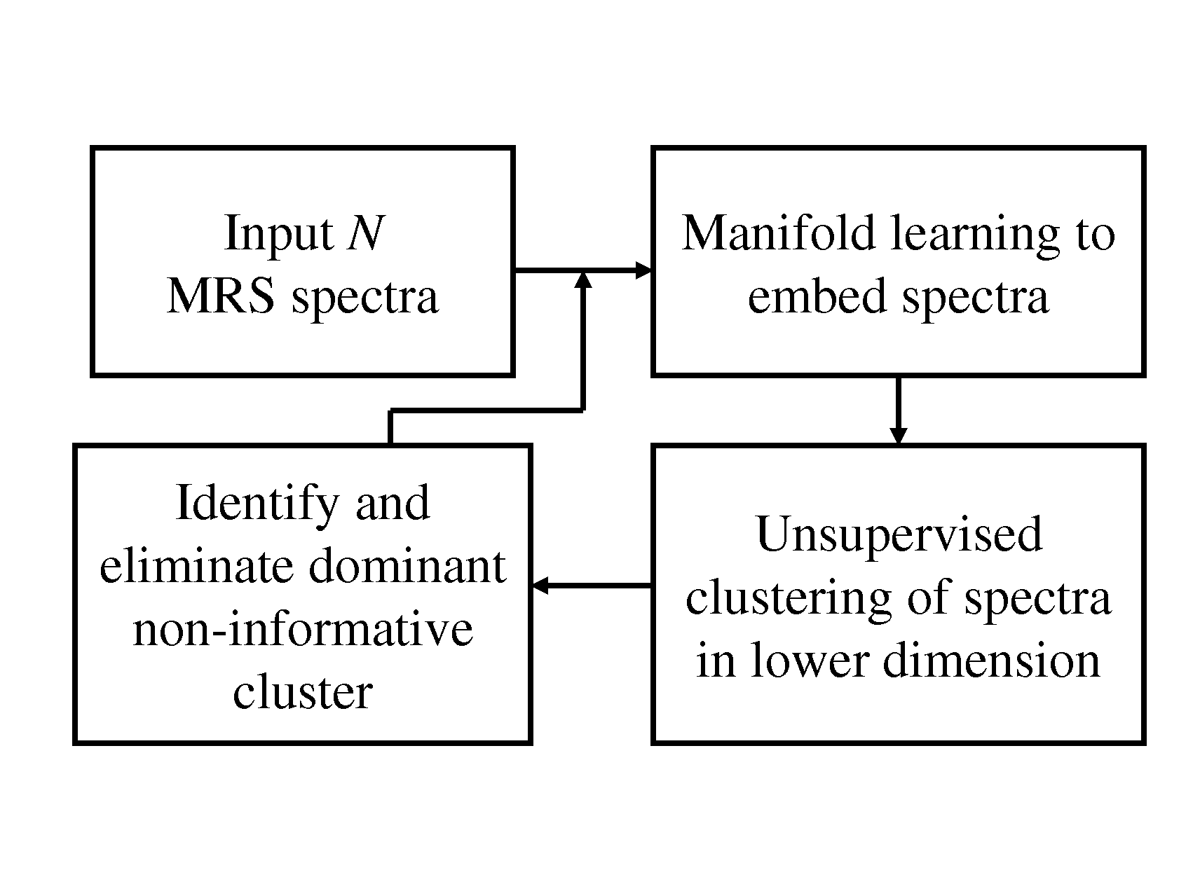
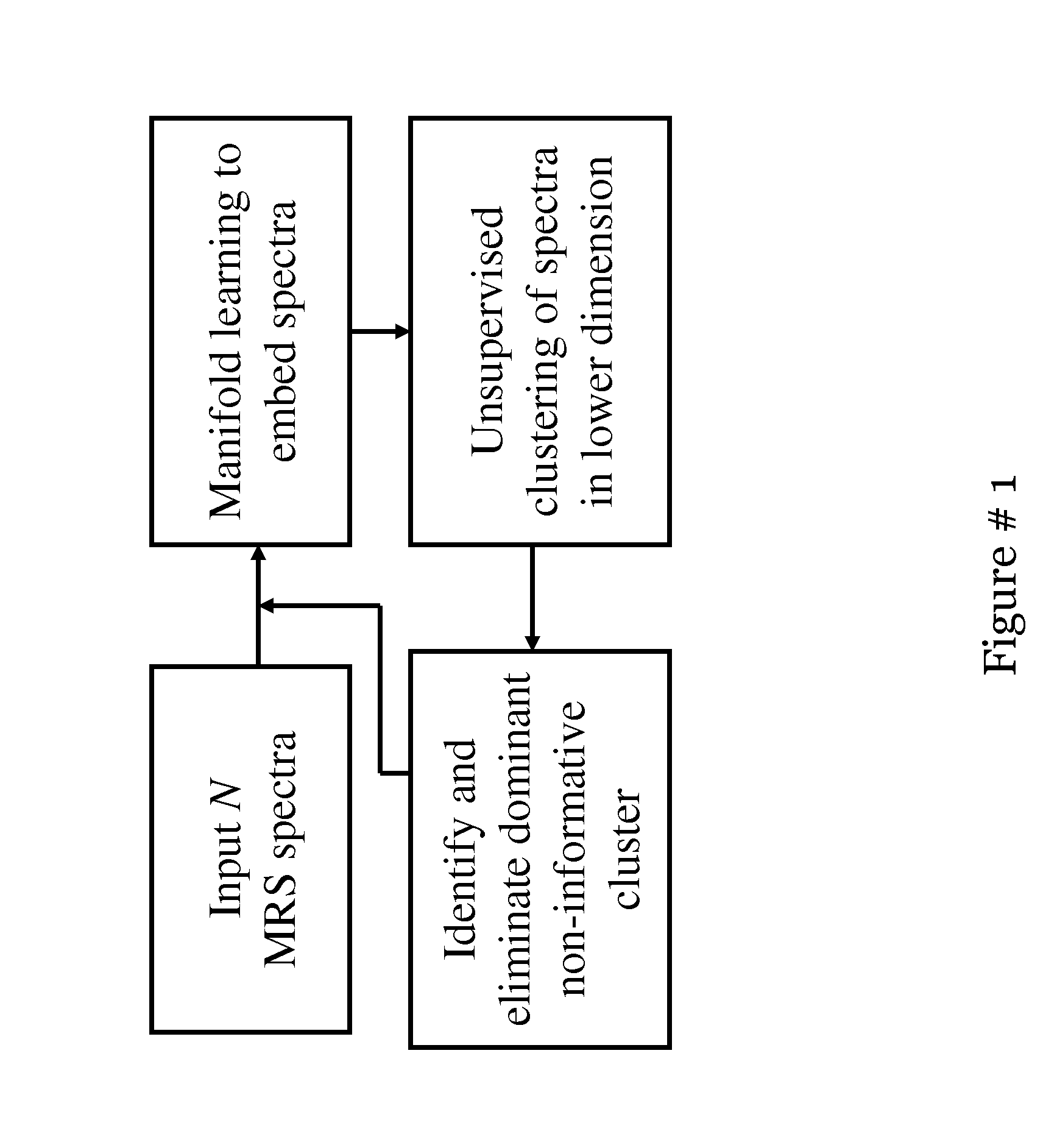
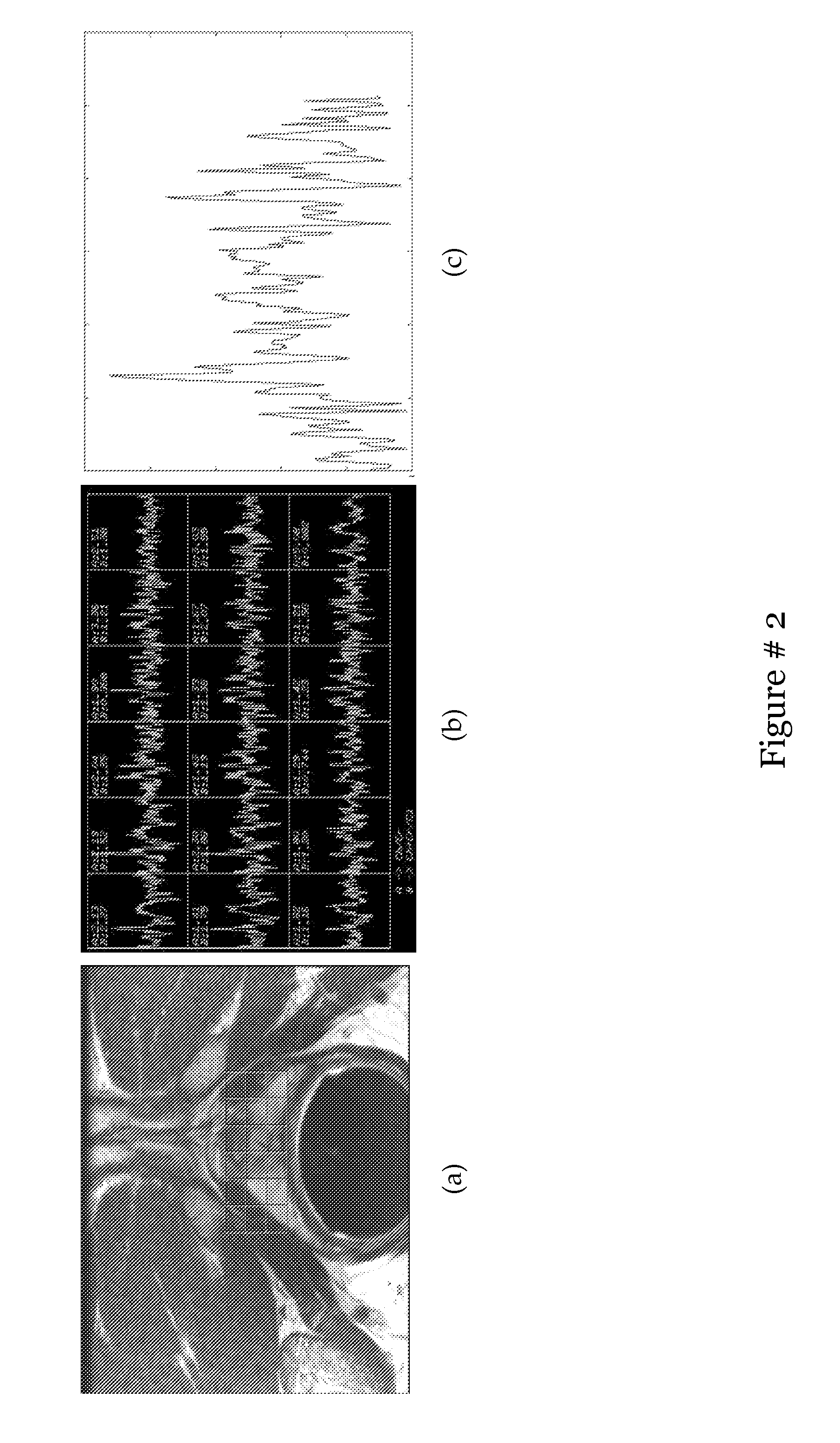
Computer assisted diagnosis (CAD) of cancer using multi-functional, multi-modal in-vivo magnetic resonance spectroscopy (MRS) and imaging (MRI)
a computer-aided diagnosis and cancer technology, applied in the field of computer-aided diagnosis and cancer classification, can solve the problems of lack of specificity, often deadly diagnosis and treatment in the late stages, and achieve the effect of reducing the dimensionality of the image feature spa
- Summary
- Abstract
- Description
- Claims
- Application Information
AI Technical Summary
Benefits of technology
Problems solved by technology
Method used
Image
Examples
example 1
A Hierarchical Unsupervised Spectral Clustering Scheme for Detection of Prostate Cancer from Magnetic Resonance Spectroscopy (MRS)
[0105]Most automated analysis work for MRS for cancer detection has focused on developing fitting techniques that yield peak areas or relative metabolic concentrations of different metabolites like choline, creatine and citrate as accurately as possible. The automated peak finding algorithms suffer from problems associated with the noisy data which worsens when a large baseline is present along with low signal to noise ratio. z-score (ratio of difference between population mean and individual score to the population standard deviation) analysis was suggested as an automated technique for quantitative assessment of 3D MRSI data for glioma. A predefined threshold value of the z score is used to classify spectra in two classes: malignant and benign. Some worked on the quantification of prostate MRSI by model based time fitting and frequency domain analysis. ...
example 2
Consensus-Locally Linear Embedding (C-LLE): Application to Prostate Cancer Detection on Magnetic Resonance Spectroscopy
[0121]Due to inherent non-linearities in biomedical data, non-linear dimensionality reduction (NLDR) schemes such as Locally Linear Embedding (LLE) have begun to be employed for data analysis and visualization. LLE attempts to preserve geodesic distances between objects, while projecting the data from the high to the low dimensional feature spaces unlike linear dimensionality reduction (DR) schemes such as Principal Component Analysis (PCA) which preserve the Euclidean distances between objects. The low dimensional data representations obtained via LLE are a function of κ, a parameter controlling the size of the neighborhood within which local linearity is assumed and used to approximate geodesic distances. Since LLE is typically used in an unsupervised context for visualizing and identifying object clusters, a priori, the optimal value of κ is not-obvious.
[0122]Row...
example 3
Multi-Modal Prostate Segmentation Scheme by Combining Spectral Clustering and Active Shape Models
[0145]In many CAD systems, segmentation of the object of interest is a necessary first step, yet segmenting the prostate from in vivo MR images is a particularly difficult task. The prostate is especially difficult to see in in vivo imagery because of poor tissue contrast on account of MRI related artifacts such as background inhomogeneity. While the identification of the prostate boundary is critical for calculating the prostate volume, for creating patient specific prostate anatomical models, and for CAD systems, accurately identifying the prostate boundaries on an MR image is a tedious task, and manual segmentation is not only laborious but also very subjective.
[0146]Previous work on automatic or semi-automatic prostate segmentation has primarily focused on TRUS images. Only a few prostate segmentation attempts for MR images currently exist. Segmenting prostate MR images was suggested...
PUM
 Abstract
Description
Claims
Application Information
 - R&D
- Intellectual Property
- Life Sciences
- Materials
- Tech Scout
- Unparalleled Data Quality
- Higher Quality Content
- 60% Fewer Hallucinations
Browse by: Latest US Patents, China's latest patents, Technical Efficacy Thesaurus, Application Domain, Technology Topic, Popular Technical Reports.
© 2025 PatSnap. All rights reserved.Legal|Privacy policy|Modern Slavery Act Transparency Statement|Sitemap|About US| Contact US: help@patsnap.com


