
Method for non-contrast enhanced pulmonary vein magnetic resonance imaging

a magnetic resonance imaging and enhanced technology, applied in the field of medical imaging, can solve the problems of limiting the diagnostic information in the image, compromising the conspicuity of the pulmonary vein, and lack of image contrast, so as to facilitate more accurate measurements of the size of the pulmonary vein ostia, suppress the signal, and enhance the conspicuity of the left atrium and the pulmonary vein.

Inactive Publication Date: 2013-08-13
BETH ISRAEL DEACONESS MEDICAL CENT INC
View PDF6 Cites 0 Cited by
  • Summary
  • Abstract
  • Description
  • Claims
  • Application Information

AI Technical Summary

Benefits of technology

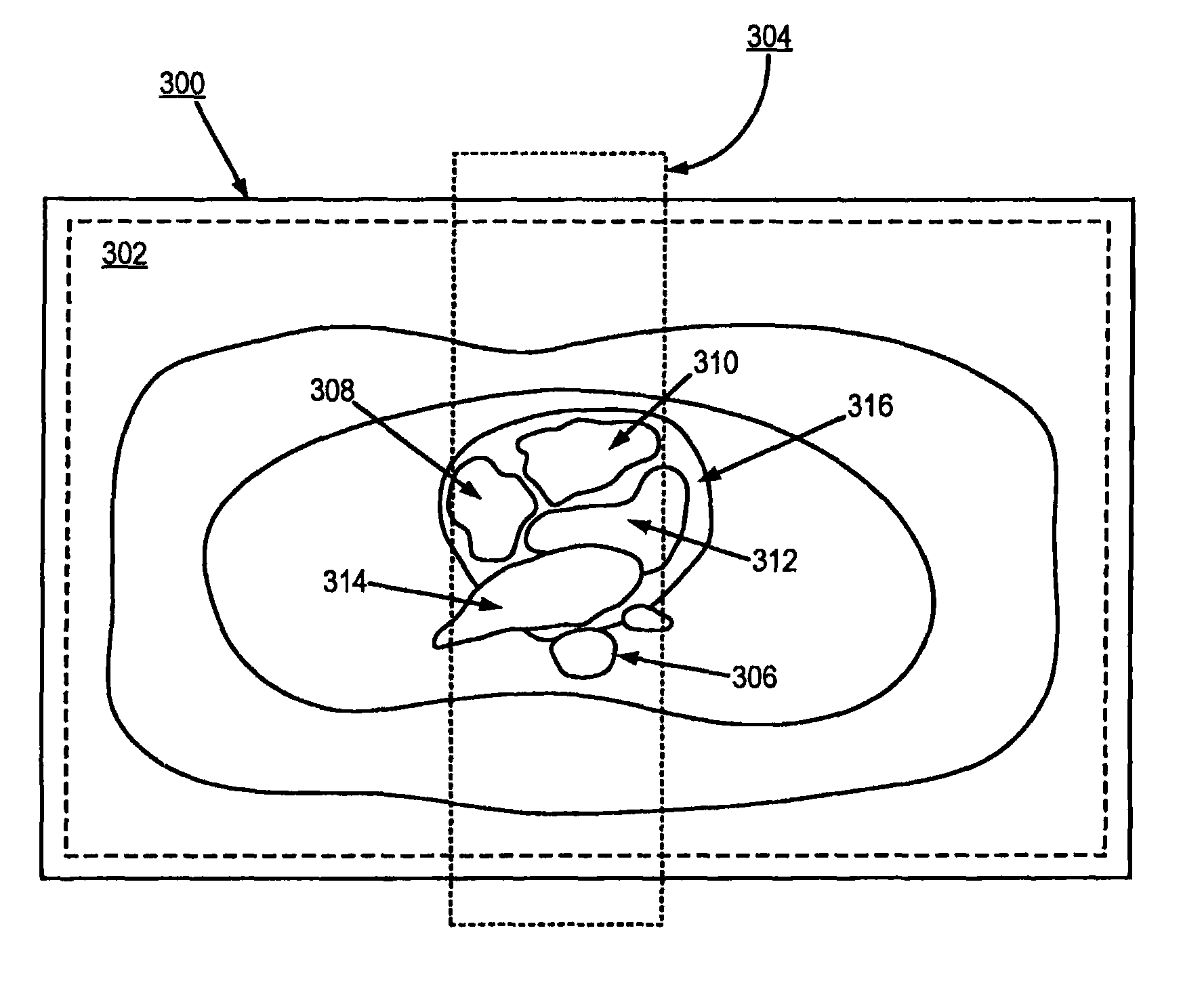
The invention provides a better way to image the veins in the lungs using MRI. By reducing the signal from the heart, the method makes it easier to see the veins and the left atrium. This improves the accuracy of measuring the size of the vein openings and the registration of MRI volumes with an ablation catheter in a procedure to isolate the veins. The method also produces three-dimensional views of the left atrium and veins without using contrast agents.

Problems solved by technology

If the central lines of k-space are acquired prior to the arrival of contrast, severe image artifacts can limit the diagnostic information in the image.
Moreover, this data acquisition typically occurs during a prolonged breath-hold by the patient, which poses difficulties for those patients with atrial fibrillation.
This lack of image contrast compromises pulmonary vein conspicuity.
Data acquisition without ECG gating also poses difficulties for imaging the pulmonary veins, since doing so results in image blurring and over-estimation of pulmonary vein size.
In subtracting these data sets, however, pulsed ASL methods can introduce phase cancellations and produce signal-to-noise ratio (SNR) loss.
Aside from the undesirable increase in imaging time required for such double-inversion methods, lack of conspicuity between pulmonary veins and the left atrium and their adjacent structures is still prevalent in pulsed ASL methods, which prevents these methods from widespread clinical use.

Method used


Image

  • Method for non-contrast enhanced pulmonary vein magnetic resonance imaging
  • Method for non-contrast enhanced pulmonary vein magnetic resonance imaging
  • Method for non-contrast enhanced pulmonary vein magnetic resonance imaging

Examples


Embodiment Construction

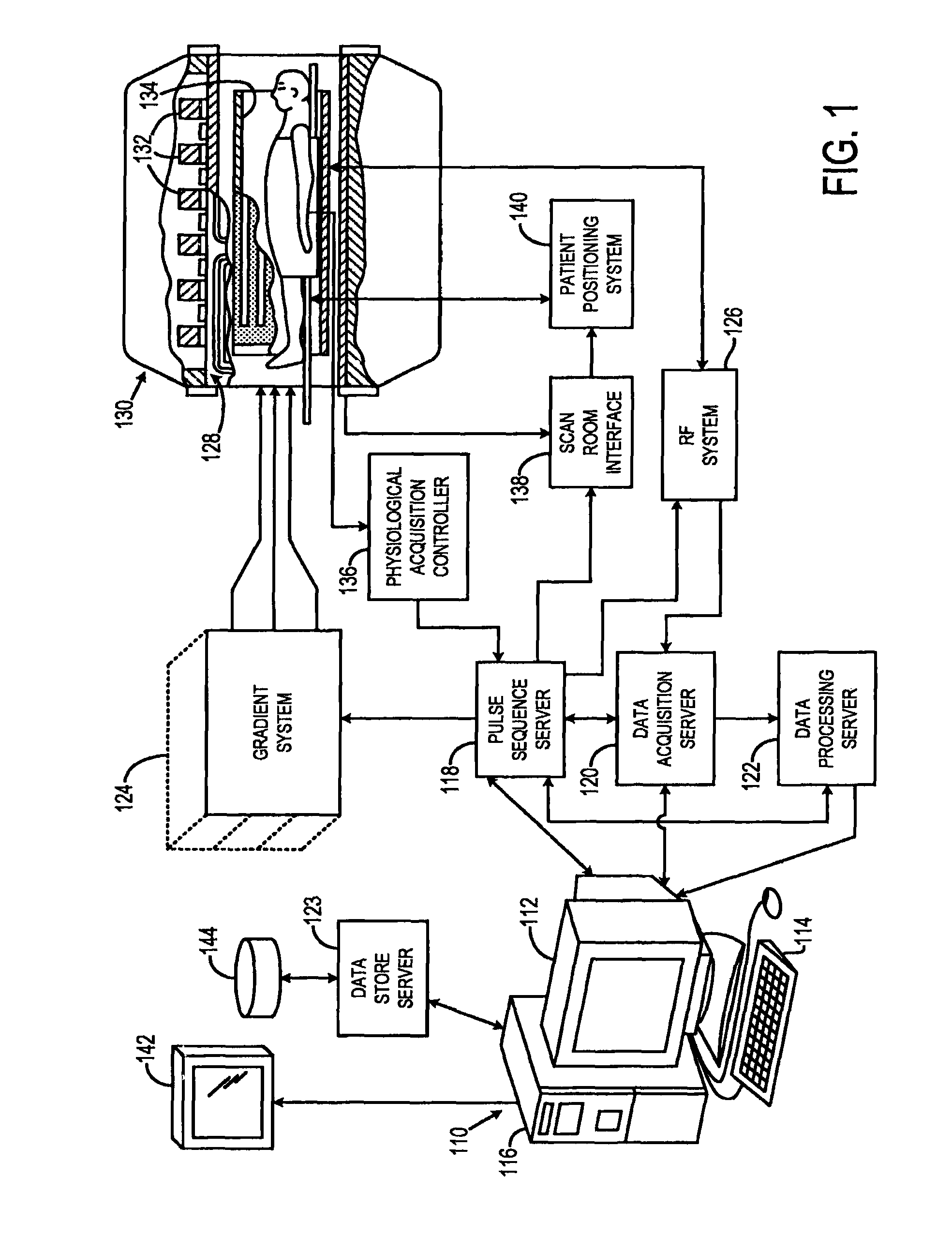
[0019]Referring particularly to FIG. 1, the present invention is employed in an MRI system. The MRI system includes a workstation 110 having a display 112 and a keyboard 114. The workstation 110 includes a processor 116 that is a commercially available programmable machine running a commercially available operating system. The workstation 110 provides the operator interface that enables scan prescriptions to be entered into the MRI system. The workstation 110 is coupled to four servers: a pulse sequence server 118; a data acquisition server 120; a data processing server 122, and a data store server 123. The workstation 110 and each server 118, 120, 122 and 123 are connected to communicate with each other.

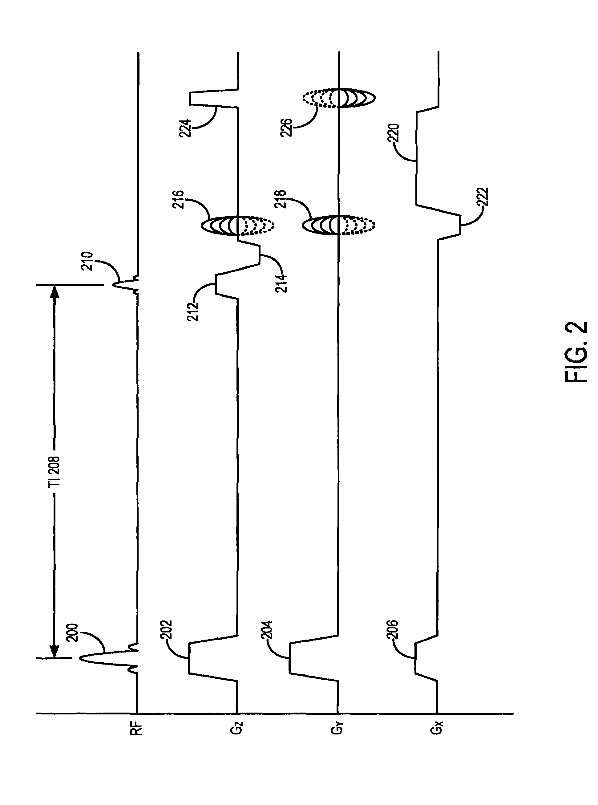
[0020]The pulse sequence server 118 functions in response to instructions downloaded from the workstation 110 to operate a gradient system 124 and an RF system 126. Gradient waveforms necessary to perform the prescribed scan are produced and applied to the gradient system 124 that e...


PUM


Abstract

A system and method for non-contrast enhanced pulmonary vein magnetic resonance imaging substantially suppresses the signal from cardiac tissue adjacent to the left atrium and pulmonary vein is provided. Significant conspicuity of the left atrium and pulmonary vein versus adjacent anatomical structures is produced. In this manner, more accurate measurements of pulmonary vein ostia size are facilitated, as well as more accurate registration of imaging volumes with a radiofrequency ablation catheter during pulmonary vein isolation procedures. In addition, more robust three-dimensional volume views of the left atrium and pulmonary vein are produced without the administration of contrast agents.

Description

CROSS-REFERENCE TO RELATED APPLICATIONS[0001]This application claims the benefit of U.S. Provisional patent application Ser. No. 61 / 229,881 filed on Jul. 30, 2009, and entitled “METHOD FOR NON-CONTRAST ENHANCED PULMONARY VEIN MAGNETIC RESONANCE IMAGING”.BACKGROUND OF THE INVENTION[0002]The present invention related to medical imaging and, more particularly, to systems and methods for non-contrast enhanced pulmonary vein MR imaging, for example, using magnetic resonance imaging (MRI).[0003]In MRI, when a substance such as human tissue is subjected to a uniform magnetic field (polarizing field B0), the individual magnetic moments of the nuclei in the tissue attempt to align with this polarizing field, but precess about it in random order at their characteristic Larmor frequency. If the substance, or tissue, is subjected to a magnetic field (excitation field B1) that is in the x-y plane and that is near the Larmor frequency, the net aligned moment, Mz, may be rotated, or “tipped”, into...

Claims


Application Information

Patent Timeline
Patent Type & Authority Patents(United States)
IPC IPC(8): A61B5/055
CPCA61B5/055G01R33/5602G01R33/5635
Inventor HU, PENGNEZAFAT, REZA
Owner BETH ISRAEL DEACONESS MEDICAL CENT INC
Learn More