

Skin permeating and cell entering (SPACE) peptides and methods of use thereof
a technology of skin permeation and cell entry, applied in the direction of fused cells, drug compositions, immunological disorders, etc., can solve the problems of poor skin penetration of macromolecules, difficult to achieve, and difficult to achieve the goal of topical sirna delivery, etc., to achieve the effect of facilitating the delivery
- Summary
- Abstract
- Description
- Claims
- Application Information
AI Technical Summary
Benefits of technology
Problems solved by technology
Method used
Image

Examples
example 1
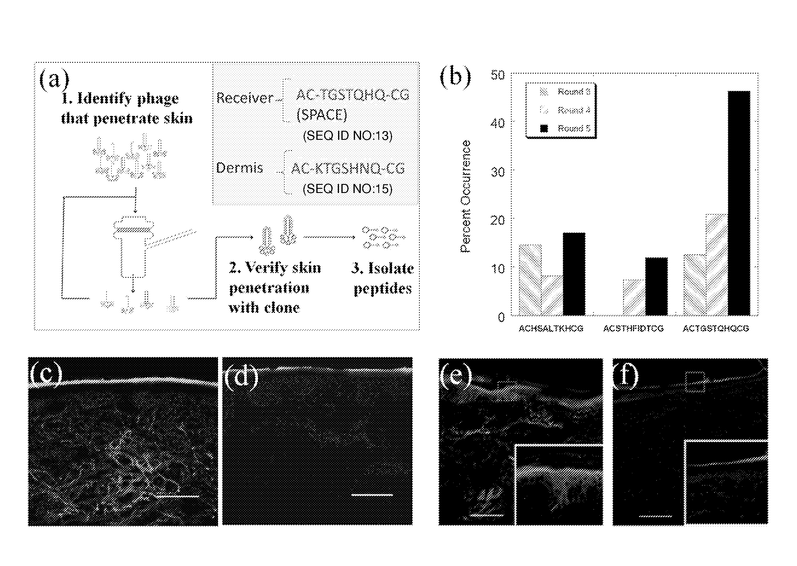
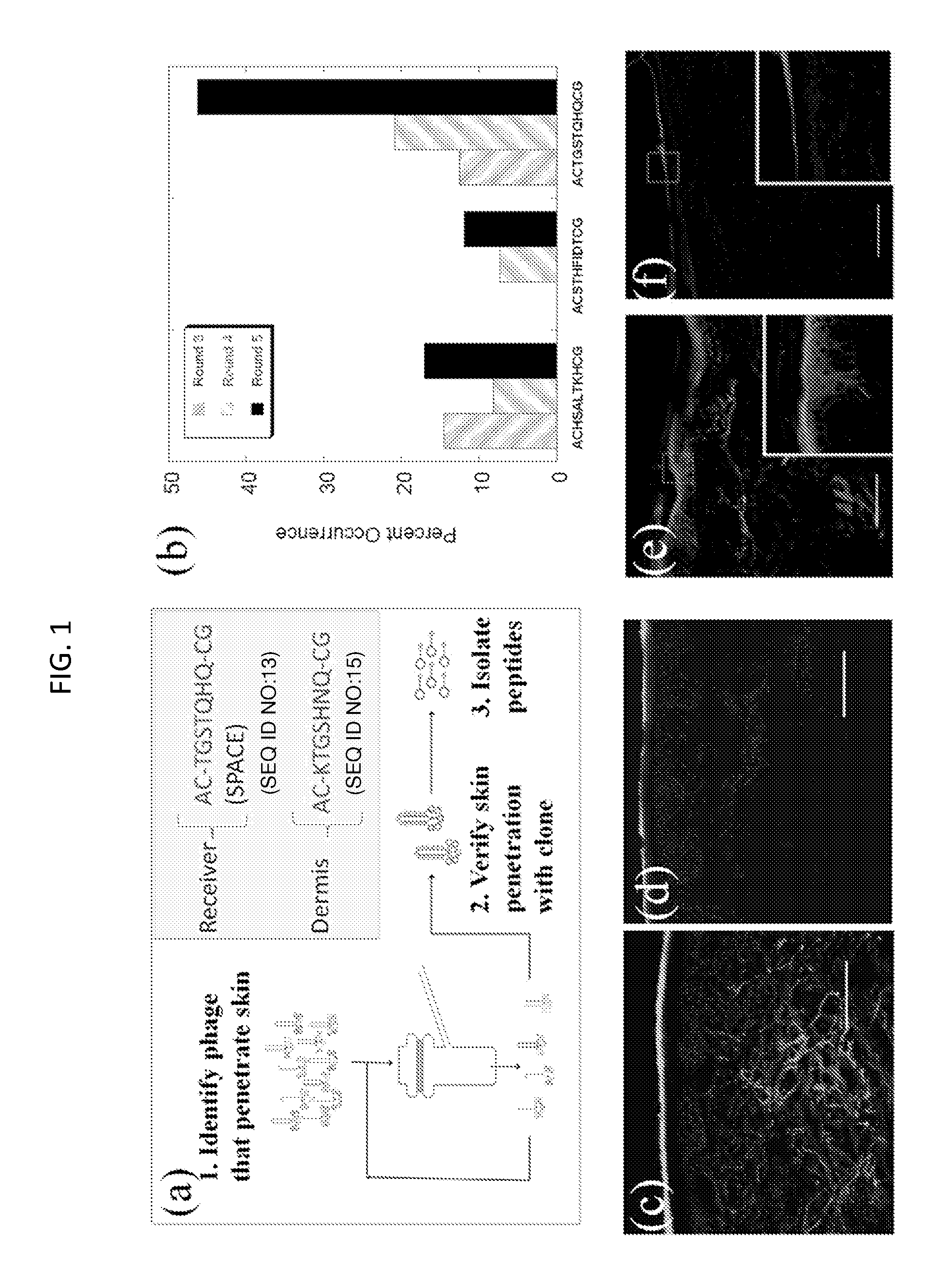
[0226]Peptides that penetrate the SC were identified using in vitro phage display as follows and as depicted generally in FIG. 1, panel A.
[0227]Phage Display
[0228]The Ph.D.-C7C Phage Display Peptide Library (New England Biolabs) was utilized. Screening studies were performed on porcine skin in Franz Diffusion cells (FDCs, Permegear). 2×10¹¹ pfu (10 μL) of phage library along with 1 mL of Phosphate Buffered Saline (PBS, pH 7.4) were placed in the donor compartment of the FDC. After 24 hours, the liquid in the receiver compartment was removed and titered by adding an aliquot of the receiver solution to 200 μL of E. Coli strain ER2738 (New England Biolabs) and plating on IPTG / Xgal plates. The number of blue plaques formed after incubation for 18 hours at 37° C. was counted and 20 plaques were randomly selected for sequencing. For subsequent screening rounds, 1 mL of the receiver solution was added to 20 mL of a 1:100 diluted overnight culture of ER2738 and grown for 4.5 hours to amplif...
example 2
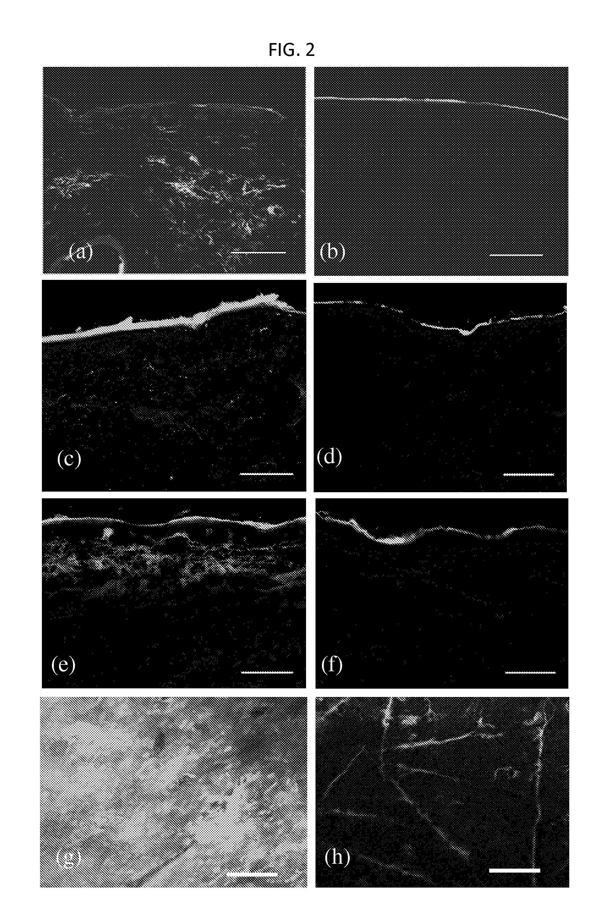
[0237]Penetration of phage across the SC is unexpected given its size (Potts R O & Guy R H (1992) Predicting skin permeability. Pharm Res 9(5):663-669; Magnusson B, Pugh W, & Roberts M (2004) Simple rules defining the potential of compounds for transdermal delivery or toxicity. Pharm Res 21(6):1047-1054; Mitragotri S (2003) Modeling skin permeability to hydrophilic and hydrophobic solutes based on four permeation pathways. J Control Release 86(1):69-92.). Large solutes (typically MW>500 Da) exhibit poor skin penetration and measurement of their transdermal permeation is often limited by the sensitivity of their detection. The M13 phage used in this study is a long filamentous particle, approximately 8 nm in width and 900 nm in length. High donor concentration (˜2×10¹¹ pfu / ml), low detection limit (˜1 pfu) and the potential for amplification facilitated assessment of dermal penetration of phage. The measured permeability of phage across porcine skin was very low; 10...
example 3
Dermis Screen
[0262]In a separate experiment, phage screening was performed to isolate phage that localized in the dermis. For the dermis screen, the phage display library was also placed in the donor compartment of the FDC. After 24 hours, the liquid from the donor compartment was removed and the skin was placed at 60° C. for 90 seconds. The epidermis was then removed from the dermis. To extract phage from the dermis, the dermis was cut up into small pieces and then homogenized (IKA disperser). The homogenate was spun down at 5,000 rpm for 5 minutes and then re-suspended in PBS and incubated at room temperature for 5 minutes. The samples were then centrifuged again and washed two more times. After the final centrifuge spin, the homogenates were re-suspended in 1% NP40 (Sigma) to elute the remaining phage from the dermis. The eluate was then plated and amplified according to the methods listed above. This was repeated for a total of 5 rounds. At the end of the fifth round of screenin...
PUM
| Property | Measurement | Unit |
|---|---|---|
| molecular weight | aaaaa | aaaaa |
| pH | aaaaa | aaaaa |
| width | aaaaa | aaaaa |
Abstract
Description
Claims
Application Information