Supercharge Your Innovation With Domain-Expert AI Agents!

Method for generating immune responses utilizing nucleic acids encoding fusion proteins comprising CD4 minimal modules and invasin polypeptides that are capable of binding to the HIV envelope and exposing cryptic neutralization epitopes

a technology of fusion proteins and nucleic acids, applied in the field of proteins, can solve the problems of general decline in the host immune system, inability to provide permanent antibodies, etc., and achieve the effect of reducing or eliminating unwanted immune responses

Inactive Publication Date: 2013-10-29
GLAXOSMITHKLINE BIOLOGICALS SA
View PDF19 Cites 1 Cited by
  • Summary
  • Abstract
  • Description
  • Claims
  • Application Information

AI Technical Summary

Benefits of technology

The present invention provides a way to make proteins that mimic the structure of a specific protein called CD4. These CD4 proteins are fused with other proteins to create a new fusion protein. This new protein structure then binds to a specific protein called Env, which causes a conformational change that makes it easier for antibodies to target the virus. This technology helps generate useful antibodies that can neutralize the virus, while reducing unwanted immune responses to other parts of the virus. The invention also includes complexes of the Env protein with the fusion protein, as well as antibodies directed against the fusion protein.

Problems solved by technology

However, it is known that antibodies directed against gp120 do not generally exhibit broad antibody responses against different HIV strains and do not induce production of neutralizing antibodies.
Furthermore, although neutralizing antibodies are typically generated in the course of HIV infection in humans, these antibodies do not provide permanent antiviral effect may in part be due to the generation of “neutralization escapes” virus mutants and to the general decline in the host immune system associated with pathogenesis.

Method used


Image

  • Method for generating immune responses utilizing nucleic acids encoding fusion proteins comprising CD4 minimal modules and invasin polypeptides that are capable of binding to the HIV envelope and exposing cryptic neutralization epitopes
  • Method for generating immune responses utilizing nucleic acids encoding fusion proteins comprising CD4 minimal modules and invasin polypeptides that are capable of binding to the HIV envelope and exposing cryptic neutralization epitopes
  • Method for generating immune responses utilizing nucleic acids encoding fusion proteins comprising CD4 minimal modules and invasin polypeptides that are capable of binding to the HIV envelope and exposing cryptic neutralization epitopes

Examples


example 1

Design of a Fusion Protein Comprising a CD4 Minimal Module

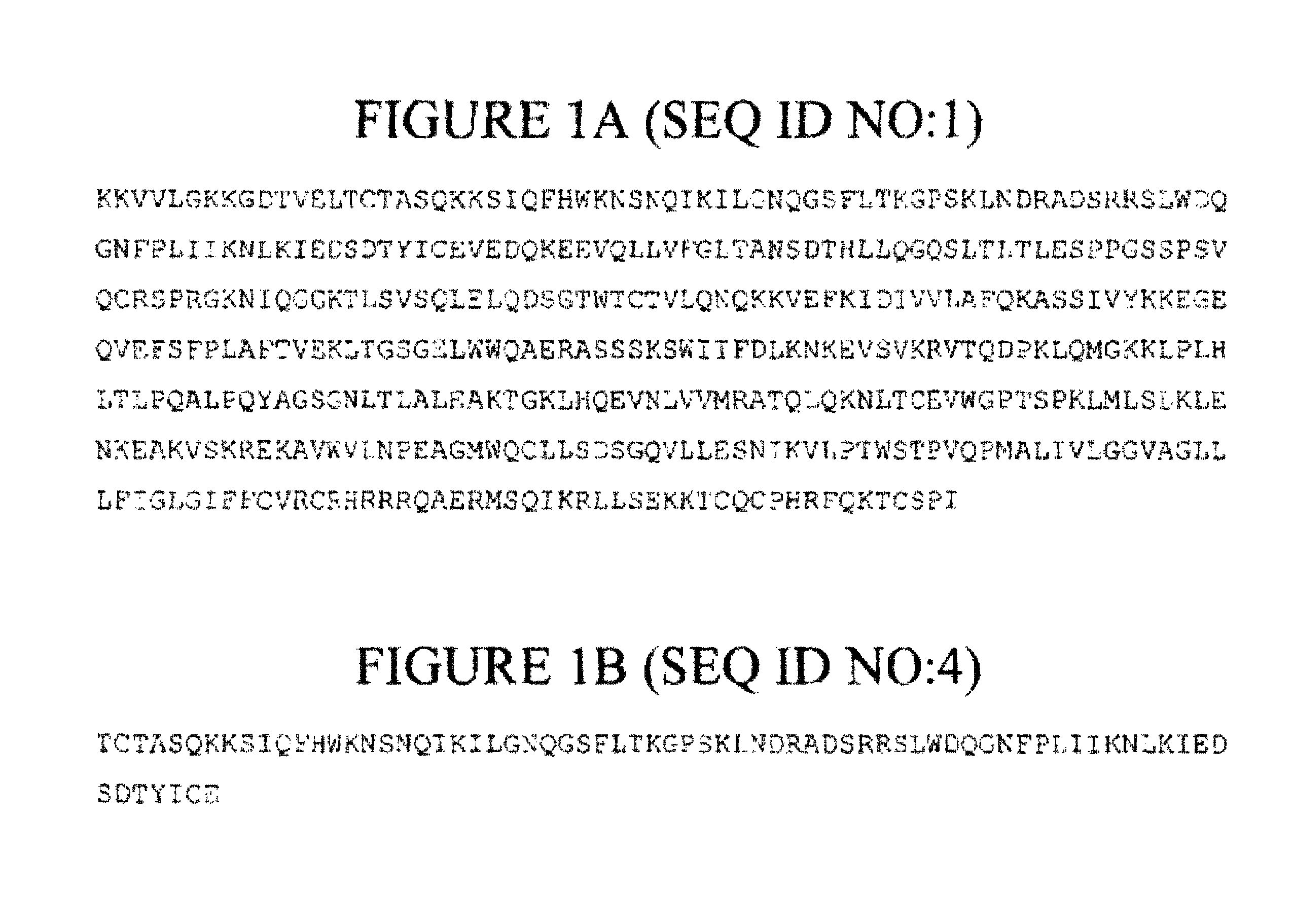
[0311]Novel fusion proteins comprising the gp120 binding region of human CD4 were designed as follows. The crystal structure of CD4-gp120 was analyzed and a region of CD4 involved in gp120 binding was identified, In particular, the region corresponding to amino acid residues 15-85 of CD4 is almost completely buried within the gp120-binding pocket (FIG. 4). This fragment was termed a CD4 minimal module and the fragment is stabilized by the presence of a disulfide bridge between Cys16 and Cys84.

[0312]Structural databases were searched for proteins having an overall configuration similar to CD4 in order to identify suitable structural scaffold into which CD4 minimal modules can be inserted to from fusion proteins. A total of 212 structures with structural similarity to native CD4 were identified. In order to minimize the risk of undesired autoimmunity, proteins from humans and primates, as well as proteins showing sequence simil...

example 2

Expression of Invasin-CD4 Fusion Proteins

[0321]Expression cassettes encoding invasin-CD4 fusion proteins shown in FIGS. 2G, 2H, 2I and 2J were expressed in E, coli. Three hours after induction with 1 mm IPTG, total cell lysate and soluble fractions were prepared and run on SDS-PAGE (4-12% MES) gels. The constructs shown in FIGS. 2H, 2I and 2J (SEQ ID NOs:11, 12 and 13, respectively) expressed proteins of the expected molecular weights in the soluble fraction.

[0322]Subsequently, expression cassettes encoding the invasin-CD4 fusion proteins shown in FIGS. 2H, 2I and 2J (SEQ ID NOs:11, 12 and 13, respectively) were expressed in bacteria (E. coli), grown to high density and inducted with 1 mM IPTG. Before induction, an uninduced sample was collected for evaluation. Following three hours of induction, cells were collected by centrifugation, lysed in lysis buffer and analyzed by SDS-PAGE / coomassie blue staining.

[0323]Proteins of the expected molecular weights were present in both induced ...

example 3

Binding Characteristics

[0325]The binding characteristics of various invasin-CD4 fusion molecules and complexes comprising these molecules were evaluated as follows.

[0326]A. Binding by CD4 Antibodies

[0327]Proteins expressed as described in Example 2 were subject to Western blotting using anti-CD4 monoclonal and polyclonal antibodies developed in-house. Briefly, different fractions of cell lysates and purified proteins (supernatant, pellet, flow through) were run on a standard SDS-PAGE gel and transferred to a nitrocellulose membrane. The members were probes with anti-CD4 monoclonals and polyclonal and the blot developed using commercially available anti-mouse and anti-rabbit IgG. Blots were scanned using Odyssey® Infrared Imaging System (LI-COR Biosciences).

[0328]The invasin-CD4 fusion proteins tested were not detectable using the anti-CD4 monoclonals and polyclonals used in these experiments.

[0329]B. Binding to gp120

[0330]Supernatants, containing the proteins expressed as described ...


PUM

PropertyMeasurementUnit
molecular weightaaaaaaaaaa
diameteraaaaaaaaaa

Abstract

Fusion proteins comprising CD4 minimal modules that bind to HIV Env polypeptides in a non-CD4 backbone are described. Also described are complexes of these fusion proteins with Env as well as methods of diagnosis, treatment and prevention using the polynucleotides and polypeptides are also provided.

Description

CROSS-REFERENCE TO RELATED APPLICATIONS[0001]This application is a divisional of U.S. application Ser. No. 11 / 597,340, filed Aug. 3, 2007, now U.S. Pat. No. 8,206,720, which is a U.S. National Stage application under 35 U.S.C. §371 of International Application PCT / US2005 / 022801 (published as WO 2006 / 085959 A2), filed Jun. 8, 2005, which claims priority to U.S. Provisional Application Ser. No. 60 / 578,151, filed Jun. 8, 2004, and U.S. Provisional Application No. 60 / 578,211, filed Jun. 8, 2004. Benefit of the filing date of each of these prior applications is hereby claimed. Each of these prior applications is hereby incorporated by reference in its entirety.[0002]This application incorporates by reference the contents of a 21.4 kb text file created on Mar. 12, 2012 and named “PAT051778DIVsequencelisting.txt,” which is the sequence listing for this application.STATEMENT OF GOVERNMENT SUPPORT[0003]The application was made with support from the United States NIAID-NIH HIVRAD under Grant ...

Claims


Application Information

Patent Timeline
Patent Type & Authority Patents(United States)
IPC IPC(8): A61K39/00C07K1/00C07H21/04A61K39/02A61K38/00A61K39/21
CPCC07K14/70514C07K2319/01A61P31/18A61P37/04
Inventor MASIGNANI, VEGASCARSELLI, MARIACAPECCHI, BARBARASHARMA, VICTORIABARNETT, SUSAN WSRIVASTAVA, INDRESH K.RAPPUOLI, RINO
Owner GLAXOSMITHKLINE BIOLOGICALS SA
Features
  • R&D
  • Intellectual Property
  • Life Sciences
  • Materials
  • Tech Scout
  • Unparalleled Data Quality
  • Higher Quality Content
  • 60% Fewer Hallucinations
Learn More