Tumour marker proteins and uses thereof

a technology of tumor markers and proteins, applied in the field of tumor markers, can solve the problems of inability to recombinantly expressed proteins, time-consuming to purify sufficient quantities of proteins, and limited use of markers

Inactive Publication Date: 2013-11-26
ONCIMMUNE
View PDF66 Cites 11 Cited by
  • Summary
  • Abstract
  • Description
  • Claims
  • Application Information

AI Technical Summary

Benefits of technology

[0040]The use of an immunoassay reagent comprising a tumour marker protein (or fragment thereof) isolated from body cavity fluids and / or excretions taken from one or more cancer patients provides significant advantages over the use of other reagents, such as recombinantly expressed or chemically synthesised polypeptides, in the clinical detection of cancer (including diagnosis, monitoring of disease recurrence or disease progression, etc).
[0045]There are further practical advantages associated with the use of ascites fluid, pleural effusion, seroma, hydrocoele or wound drainage fluid, as a source of tumour marker proteins. These fluids may be readily removed from patients in relatively large volumes as part of the therapeutic strategy. This material, which would otherwise be discarded, is a valuable source of useful assay reagent.
[0046]Given that fluids such as ascites fluid, pleural effusion, seroma, hydrocoele or wound drainage fluid are produced in large volumes, there was doubt as to whether the concentration of tumour marker proteins in such fluids would be high enough to enable such fluids to be used as a practical source of antigens. One might reasonably expect the concentration of tumour marker proteins to be more dilute in such fluids as compared to blood or serum. Surprisingly, the inventors observed that the concentrations of tumour marker proteins in such fluids are in fact significantly higher than in serum. Accordingly, there are substantial benefits to be gained in terms of yield in recovering tumour marker proteins from such fluids.
[0047]Furthermore, it has also been observed by the inventors that additional significant advantages can be secured by pooling body cavity fluid samples or excretions from two or more patients. Apart from increasing protein yield, the product secures at least as good a detection rate as marker protein from an individual sample while, at the same time, being more consistent in its characteristics from batch to batch. Thus, adequate affinity of the antigen can be relied upon every time.
[0048]In particular embodiments the methods of the invention may comprise immunoassays to (simultaneously) detect two or more types of autoantibodies, each having specificity for different tumour marker proteins or for different epitopes on the same tumour marker proteins. These methods will typically involve use of a panel of two or more assay reagents, each reagent comprising a different tumour marker protein. These methods, which may be hereinafter referred to as “panel assays”, utilise a panel of two or more reagents to monitor the overall immune response of an individual to a tumour or other carcinogenic / neoplastic change. These methods thus detect a “profile” of the immune response in a given individual, indicating which tumour markers elicit an immune response resulting in autoantibody production. The use of a panel of two or more reagents to monitor production of autoantibodies against two or more different tumour markers is generally more sensitive than the detection of autoantibodies to single markers and gives a much lower frequency of false negative results.

Problems solved by technology

These markers are of limited use earlier in the course of the disease, for example in early detection or in the screening of asymptomatic patients.
In addition, the desired protein is generally present at low concentration, therefore it is time-consuming to purify sufficient quantities of protein.
In addition, refolding of recombinantly expressed proteins may not be appropriate, thus giving an incorrect conformation for auto-antibody recognition.
Tumour tissue is usually only available in small quantities and the purification of proteins therefrom is laborious and time consuming.
Serum samples are usually available only in small quantities, therefore it is difficult to purify sufficient quantities of protein.

Method used


Image

  • Tumour marker proteins and uses thereof
  • Tumour marker proteins and uses thereof
  • Tumour marker proteins and uses thereof

Examples


example 1

General Protocol for Purification of MUC1 Antigen

[0093]Monoclonal anti-MUC1 antibody B55 (also known as NCRC 11, Xoma Corporation) is conjugated to CNBr-sepharose beads. Other anti-MUC1 monoclonal antibodies may be substituted for B55.

[0094]Tumour-induced body fluids (e.g. pleural effusion, ascites, seroma or wound drainage fluid) are diluted 1 / 10 with phosphate buffered saline (PBS) and filtered to 0.45 μm.

[0095]Diluted body fluids are incubated with the anti-MUC1 sepharose beads (25 ml diluted fluid to 1 ml packed volume of beads) overnight at 4° C. with rolling (“batch” method) or re-circulated overnight through a packed column containing anti-MUC1 sepharose beads (“column” method).

“Batch” Method:

[0096]Beads are packed by centrifugation and the supernatant removed;

[0097]Beads re-suspended in 5-10 ml PBS and rolled for 10 mins then packed by centrifugation and the supernatant removed; repeat 5 times (or until A280 nm˜0)

[0098]Beads re-suspended in 5 ml 100 mM DEA pH 11, and rolled ...

example 2a

General Protocol for Purification of MUC16 Antigen (Previously Known as CA125)

[0110]One volume (e.g. 50 ml) of saturated ammonium sulphate was added to one volume (e.g. 50 ml) of tumour-induced body fluid (e.g. pleural effusion, ascites, seroma or wound drainage fluid) and incubated overnight at 4° C.

[0111]The resultant precipitate is collected by centrifugation (3500 rpm for 30 min in a standard benchtop centrifuge) and resuspended in ½ volume PBS.

[0112]This resuspension is subjected to gel filtration chromatography through an S300 column (2.5×100 cm) using PBS as the eluting buffer.

[0113]Fractions (5 or 10 ml) are collected and assayed by ELISA for MUC16, using for instance anti-CA125 from ICN or the anti-MUC16 antibody VK8 (Memorial Sloane Kettering, New York), prior to pooling MUC16 positive fractions and storage at −20° C.

[0114]In order to remove contaminating immunoglobulins, MUC16 pools are incubated with NaSCN (to 1.5M) for 10 mins, DTT (to 50 mM) for 30 mins, then iodoaceta...

example 2b

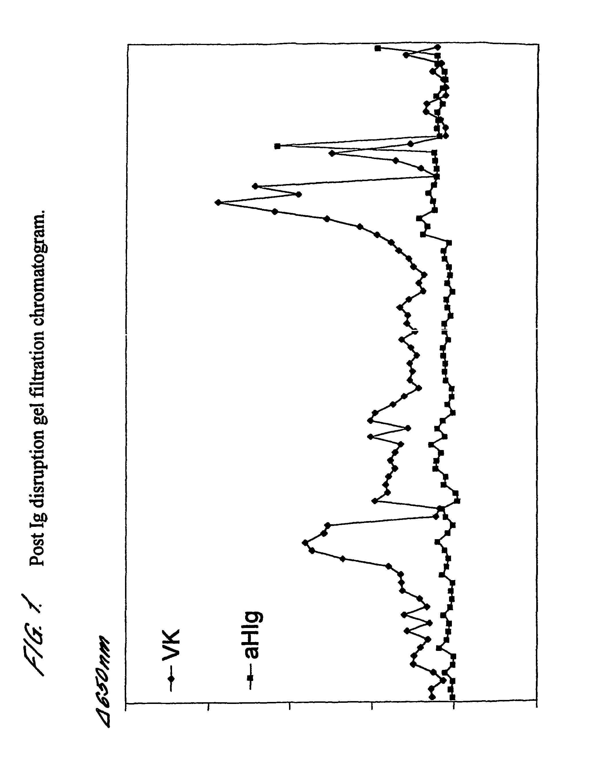
Post Ig Disruption Gel Filtration Chromatography

[0117]For a sample prepared in the manner described above, fractions from a post-Ig disruption gel filtration were assayed for MUC16 using anti-MUC16 antibody VK8 and for human Ig using an anti-human Ig. The results are shown in FIG. 1. As is clearly demonstrated, two substantially immunoglobulin free MUC16 peaks are eluted.


PUM

PropertyMeasurementUnit
volumeaaaaaaaaaa
pHaaaaaaaaaa
volumeaaaaaaaaaa

Abstract

The invention relates to tumor marker proteins and their preparation from fluids from one or more cancer patients, wherein said fluids are those which collect in a body cavity or space which is naturally occurring or which is the result of cancer or medical intervention for cancer. The invention also relates to preparation of tumor marker proteins from excretions taken from patients with cancer. The tumor marker proteins are useful as immunoassay reagents in the detection of cancer-associated anti-tumor marker autoantibodies.

Description

CROSS REFERENCE TO RELATED APPLICATIONS[0001]This application is the U.S. national phase of International Application No. PCT / GB2003 / 004950 filed on Nov. 13, 2003 and published in English on May 27, 2004 as International Publication No. WO 2004 / 044950, which application claims priority to GB Patent Application No. 0226622.9 filed on Nov. 14, 2002, the contents of which are incorporated by reference herein.FIELD OF THE INVENTION[0002]The invention relates to tumour marker proteins and their preparation from fluids from one or more cancer patients, wherein said fluids are those which collect in a body cavity or space which is naturally occurring or which is the result of cancer or medical intervention for cancer. Exemplary fluids are ascites, pleural effusion, seroma, hydrocoele and wound drainage fluid. The invention also relates to preparation of tumour marker proteins from excretions taken from patients with cancer.[0003]The said tumour marker proteins are useful in cancer detectio...

Claims


Application Information

Patent Timeline
Patent Type & Authority Patents(United States)
IPC IPC(8): G01N33/574A61P35/00G01N33/564
CPCG01N33/564G01N33/57488C07K1/36G01N33/6854G01N2800/24A61P35/00C07K14/4748G01N33/574G01N33/57473G01N33/57484
Inventor ROBERTSON, JOHN FORSYTH RUSSELLGRAVES, CATHERINE ROSAMUND LOUISE
Owner ONCIMMUNE
Why Eureka
Try Eureka