Apparatus for detecting the position of a percutaneously-inserted intravenous catheter

a technology for apparatuses, which is applied in the field of apparatus for detecting the position of percutaneously inserted intravenous catheters, can solve the problems of clinical deterioration, inability to insert further, and inability to use a procedure which involves taking x-ray images of neonates

Active Publication Date: 2013-12-31
NEWCASTLE UPON TYNE HOSPITALS NAT HEALTH SERVICE TRUST
View PDF7 Cites 58 Cited by
  • Summary
  • Abstract
  • Description
  • Claims
  • Application Information

AI Technical Summary

Problems solved by technology

However, if the catheter has not been inserted sufficiently far it cannot be inserted further due to loss of the sterile insertion field when performing the X-ray.
Using a procedure which involves taking X-ray images of a neonate is not desirable for a number of reasons.
First, exposure to X-rays can itself lead to harm.
Secondly, it is necessary to disturb or move the neonate from one location to another to perform the X-ray.
Such movement of an ill neonate can cause distress leading to clinical deterioration, and also may involve delay.
Thirdly, the very fine gauge intravenous lines used in neonatal practice may be difficult to localise accurately on X-ray, even with the use of contrast media.
A problem associated with using the connectors of the Vygon® type is that noise arising from interference, typically mains interference and movement artifacts, prevents an accurate pattern being determined.
Excessive noise is produced due to the very high impedance of electrically conductive saline resulting from the very small diameter of the catheter.
However, increasing the strength of the saline solution can damage the red blood cells and carries risk of hypernatremia.
Neubauer recognizes that over-supplying sodium and chloride can be harmful to neonates and overcomes this in the smallest of children by first filling the catheter with glucose solution which may be sprayed out of the catheter without any risk in the event of difficulty in inserting the catheter being encountered.
As recognized by Neubauer, when filled with physiological saline the impedance of such catheters is very high and the electrical signal detected by such a catheter is accompanied by a great deal of noise due to this high impedance.

Method used


Image

  • Apparatus for detecting the position of a percutaneously-inserted intravenous catheter
  • Apparatus for detecting the position of a percutaneously-inserted intravenous catheter
  • Apparatus for detecting the position of a percutaneously-inserted intravenous catheter

Examples


Embodiment Construction

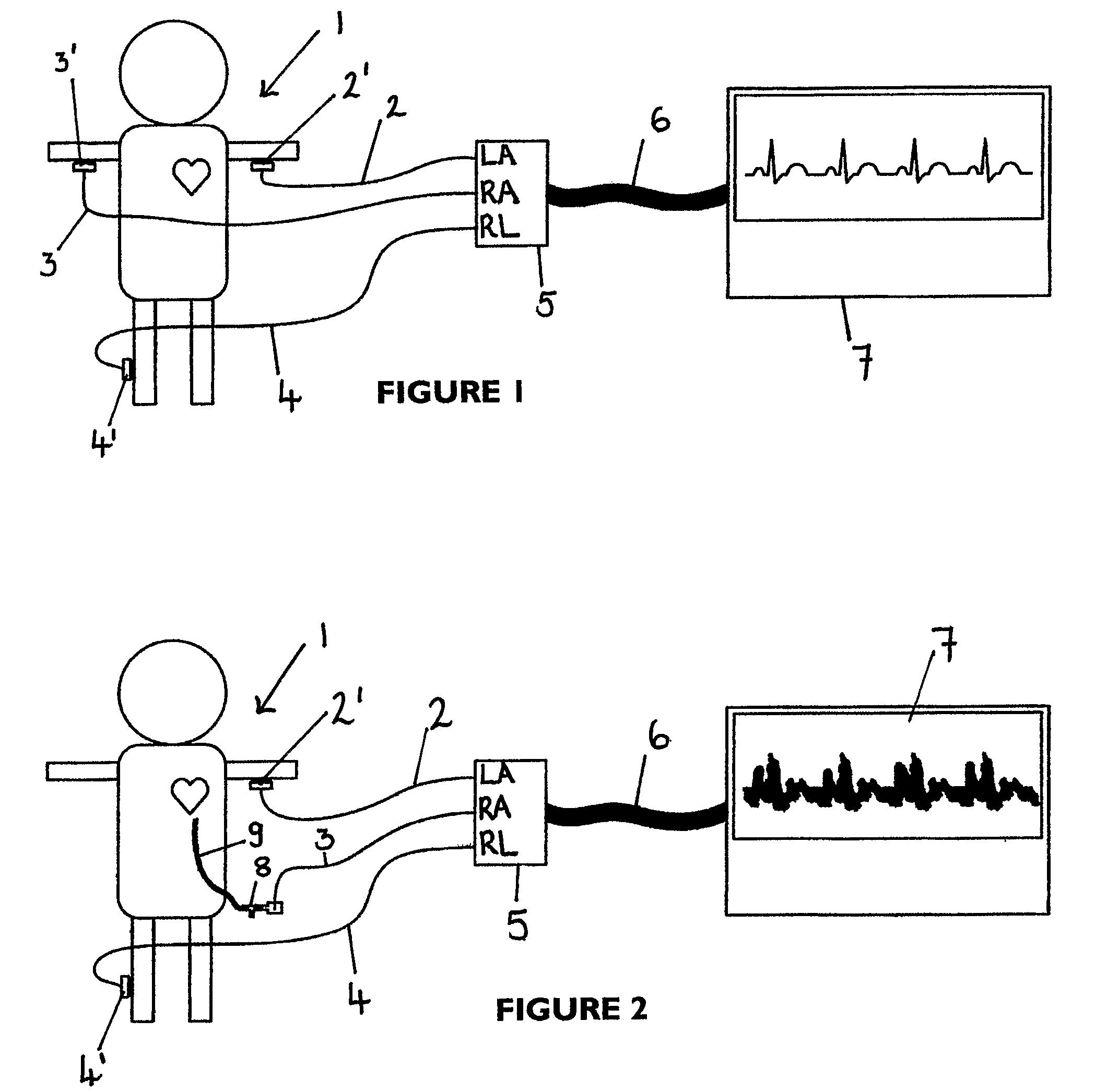
[0033]The ensuing description assumes that the ECG monitor is configured as ECG Lead I (left arm-right arm) (Einthoven system) using right leg as reference. Other lead configurations are equally possible, such as configurations in which the left leg is used as a reference.

[0034]Referring now to FIG. 1, there is shown a typical arrangement of an ECG on an individual 1 in which leads 2, 3 and 4 are connected to the left arm, right arm and right leg respectively by electrodes 2′, 3′ and 4′. The leads 2 to 4 are connected respectively to the left arm (LA), right arm (RA) and right leg (RL) terminals on a patient lead connector 5, which is itself connected to by a cable 6 to an ECG monitor 7. Interference is not a significant problem with this arrangement as each of the leads 2 to 4 presents substantially the same, relatively low impedance.

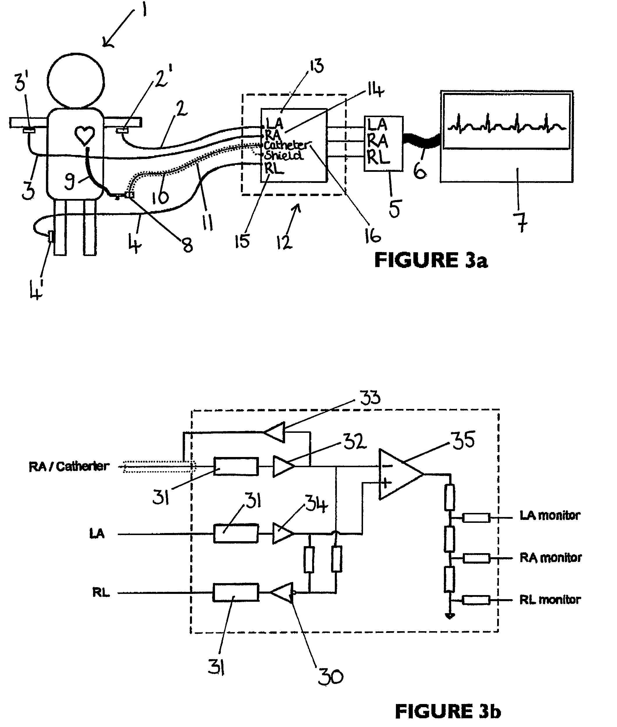
[0035]Referring now to FIG. 2, where the individual 1 is a neonate, instead of the lead 3 which connects to the RA terminal of the patient lead connec...


PUM


Abstract

An apparatus that is able to detect the position of a catheter. The apparatus utilizes a catheter filled with electrically-conductive physiological saline and a connector for establishing an electrical connection between the saline of the catheter and an input of a controller. The controller includes at least one output connectable to a standard ECG lead connector, wherein the controller includes circuitry for generating a low impedance output signal. The controller provides an output which replicates the input a standard ECG patient lead connector is configured to receive. The apparatus provides a more convenient and cost effective solution for providing specialized ECG functions without having to replace a hospital's existing ECG beside monitoring equipment.

Description

FIELD OF THE INVENTION[0001]The present invention relates to an apparatus for detecting the position of a percutaneously-inserted intravenous catheter and in particular to such an apparatus for use in neonates and in adults when a catheter is required to be positioned close to the heart.BACKGROUND OF THE INVENTION[0002]Percutaneously-inserted intravenous catheters are used in neonates for a number of reasons including parenteral nutrition, infusing drugs and taking blood for example. When carrying out such procedures the end of the catheter is inserted into a vein and positioned in close proximity to the heart in the vena cava. In some procedures performed shortly after birth, an umbilical vein is used.[0003]The current procedure for inserting a catheter uses a catheter marked with graduations. To insert the catheter marked with graduations into a neonate involves measuring the baby and applying a formula to determine to which graduation the catheter should be inserted and then taki...

Claims


Application Information

Patent Timeline
Patent Type & Authority Patents(United States)
IPC IPC(8): A61B5/0402A61B5/308
CPCA61B5/0428A61B5/042A61B5/283A61B5/30
Inventor GRIFFITHS, CLIVEWHITAKER, MIKE
Owner NEWCASTLE UPON TYNE HOSPITALS NAT HEALTH SERVICE TRUST
Why Eureka
Try Eureka