Cranial evacuation system and use thereof

a technology of cranial evacuation and cranial nerve, which is applied in the field of cranial evacuation system, can solve the problems of difficult to maintain visual and physical contact of solid matter in a working cavity, and difficulty in visualizing the cavity, so as to facilitate brain tissue displacement, stop bleeding, and expose solid matter

Active Publication Date: 2014-03-25
ABRAHAMS JOHN M
View PDF18 Cites 18 Cited by
  • Summary
  • Abstract
  • Description
  • Claims
  • Application Information

AI Technical Summary

Benefits of technology

[0007]In one aspect, the invention provides a method of removing solid matter (for example, tumor tissue, a blood clot, a cyst, a brain lesion, or infected brain tissue (e.g., an abscess or a bacterial or fungal infection)) disposed within brain tissue of a subject, and then controlling bleeding once the solid matter has been removed. The method involves securing a cranial anchor to an opening in a skull of the subject, where the anchor defines an anchor passage running therethrough. Then a channel member is passed through the anchor passage of the anchor to facilitate displacement of brain tissue and to expose the solid matter. The channel member has a first end and a second end and defines a working channel passing through the channel member from the first end to the second end. Optionally, a removable, solid trocar can be inserted into the working channel to displace the brain tissue and to expose the solid matter. The trocar, if used, then is removed to leave the working channel open for subsequent manipulations by the surgeon. The solid matter is removed via the working channel to create a cavity where the solid matter used to reside. In other words, the cavity is defined by the brain tissue that surrounded the solid matter prior to removal of the solid matter. A flowable hemostat is introduced into the cavity via the working channel to contact the brain tissue that surrounded the solid matter prior to its removal. An inflatable balloon introduced via the working channel is inflated such that the wall of the inflated balloon compresses the hemostat against the brain tissue that surrounded the solid matter prior to its removal thereby to stop the bleeding from blood vessels disposed within the brain tissue surrounding the cavity.
[0009]The anchor passage may have an internal diameter of from 1 mm to 100 mm, from 2 mm to 80 mm, from 3 mm to 75 mm, from 4 mm to 65 mm, from 5 mm to 50 mm, from 6 mm to 45 mm, 7 mm to 40 mm, from 8 mm to 35 mm, or from 10 mm to 30 mm. Furthermore, if desired, the channel member can further include a removable, solid trocar disposed within the working channel. The trocar can further facilitate the displacement of brain tissue to expose the solid matter. When a trocar is used, it is removed from the working channel to permit the surgeon to remove the solid matter via the working channel. The working channel can be from 2 cm to 15 cm long, from 3 cm to 12 cm long, from 4 cm to 11 cm long, or from 5 cm to 10 cm long, and can have an internal diameter of between 7 mm and 40 mm, or between 10 mm and 30 mm. The trocar, when used, is dimensioned to be inserted within the working channel.
[0014]The catheter can be a dual lumen catheter that permits the introduction of the hemostat via a first lumen and the introduction of the inflatable balloon via a second lumen. Optionally, the catheter can include an actuator that facilitates introduction of the inflatable balloon into the subject, and / or an actuator for inflating the balloon within the subject, and / or an actuator for introducing the hemostat into the subject.

Problems solved by technology

Given the fluid, non-self supporting and pliable nature of brain tissue, especially in the brain cortex, one of the problems associated with this type of surgery is that, once the solid matter has been exposed it can be difficult to maintain visual and physical contact of the solid matter in a working cavity because the brain tissue naturally moves or flows into the working cavity and re-covers the solid matter.
Furthermore, once the solid matter has been removed, the brain tissue surrounding the cavity created by resection of the solid matter typically tries to flow into the cavity, making it difficult to visualize the cavity (similar to looking into a collapsed bag or balloon).
Keeping both the working cavity and the cavity created by removal of the solid matter open for manipulation and visible, especially when trying to identify the source of bleeding, can be difficult if the surgeon is operating without an assistant.
Another problem associated with the surgery is stopping bleeding that occurs once the solid matter has been removed.

Method used


Image

  • Cranial evacuation system and use thereof
  • Cranial evacuation system and use thereof
  • Cranial evacuation system and use thereof

Examples


Embodiment Construction

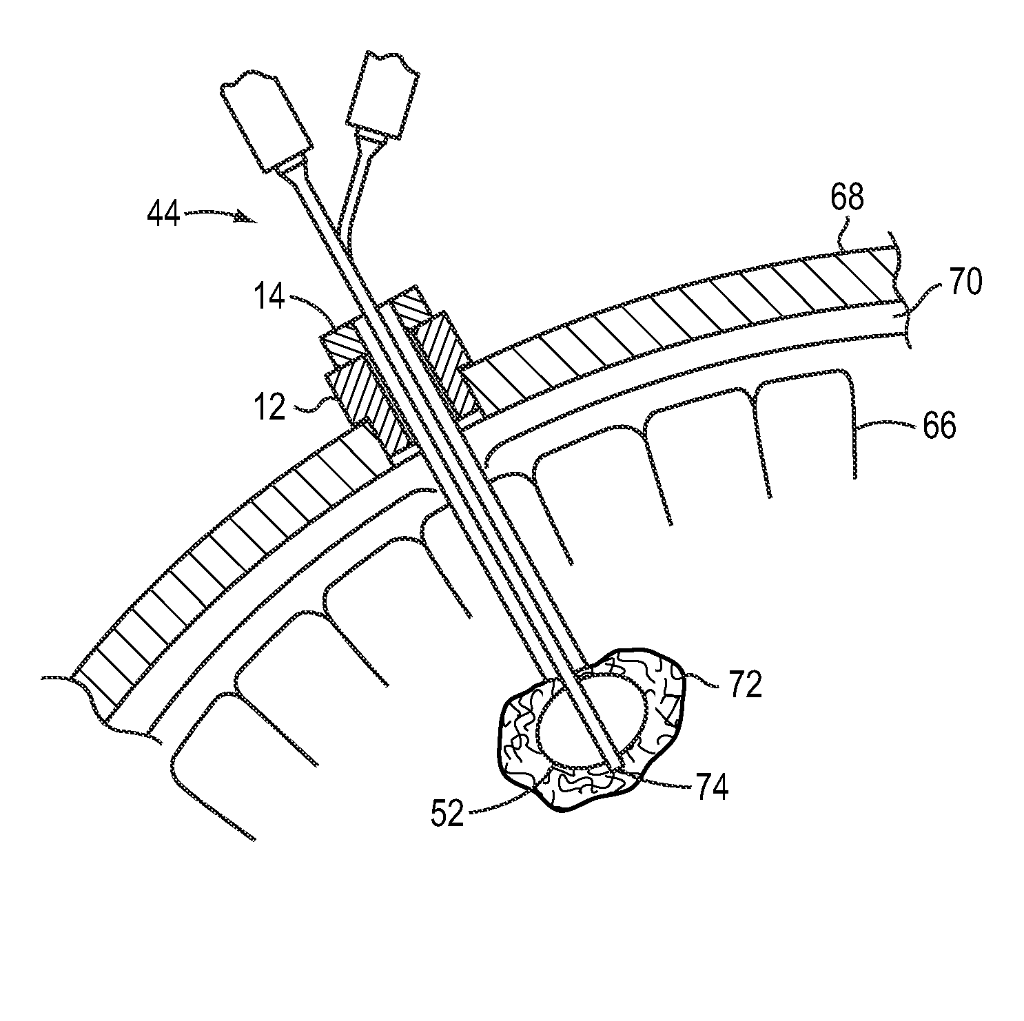
[0025]The invention relates to a method and device or system for facilitating the removal of undesirable solid matter from brain tissue, and for controlling intracranial bleeding in the region of the brain tissue once the solid matter has been removed.

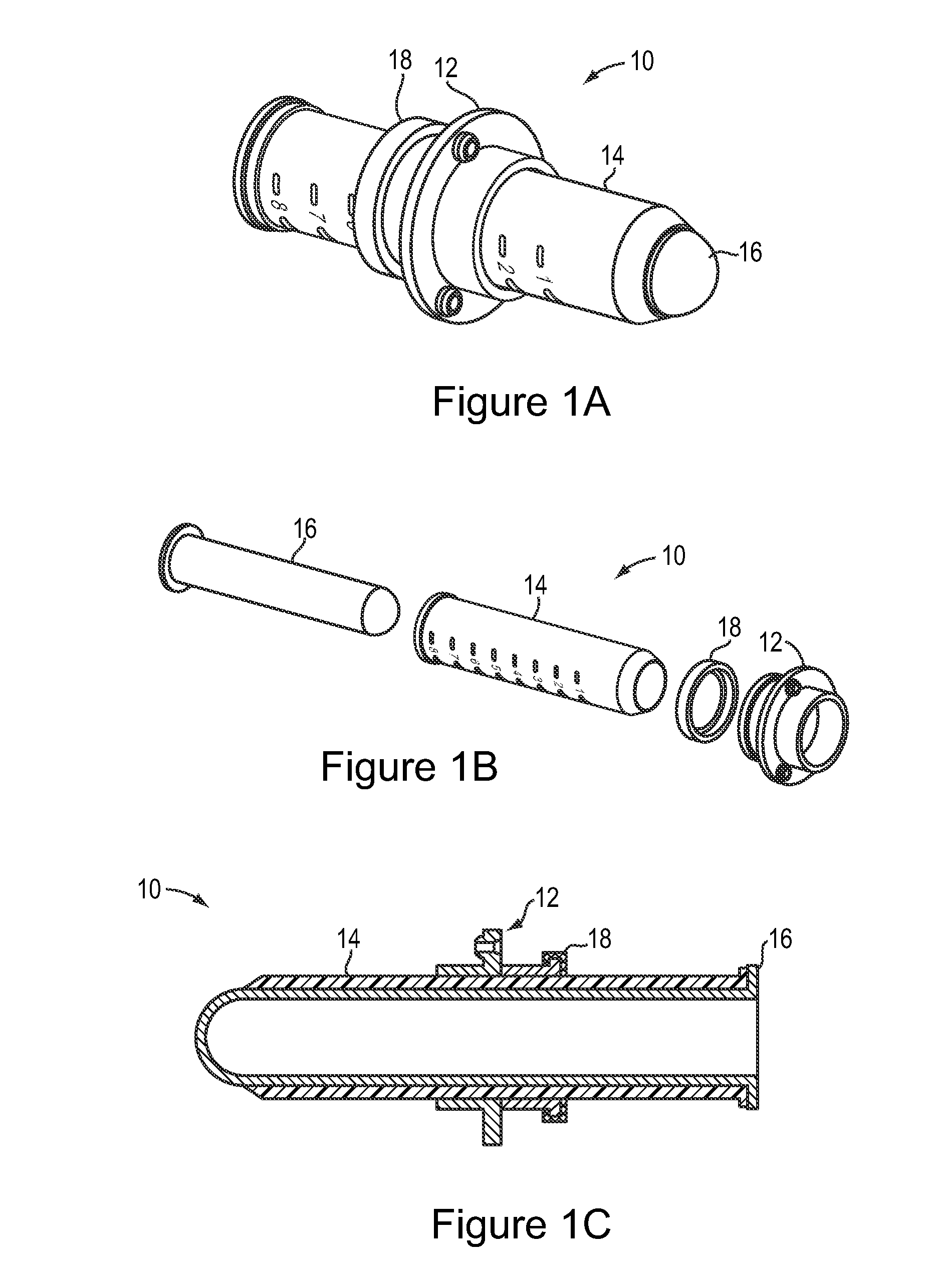
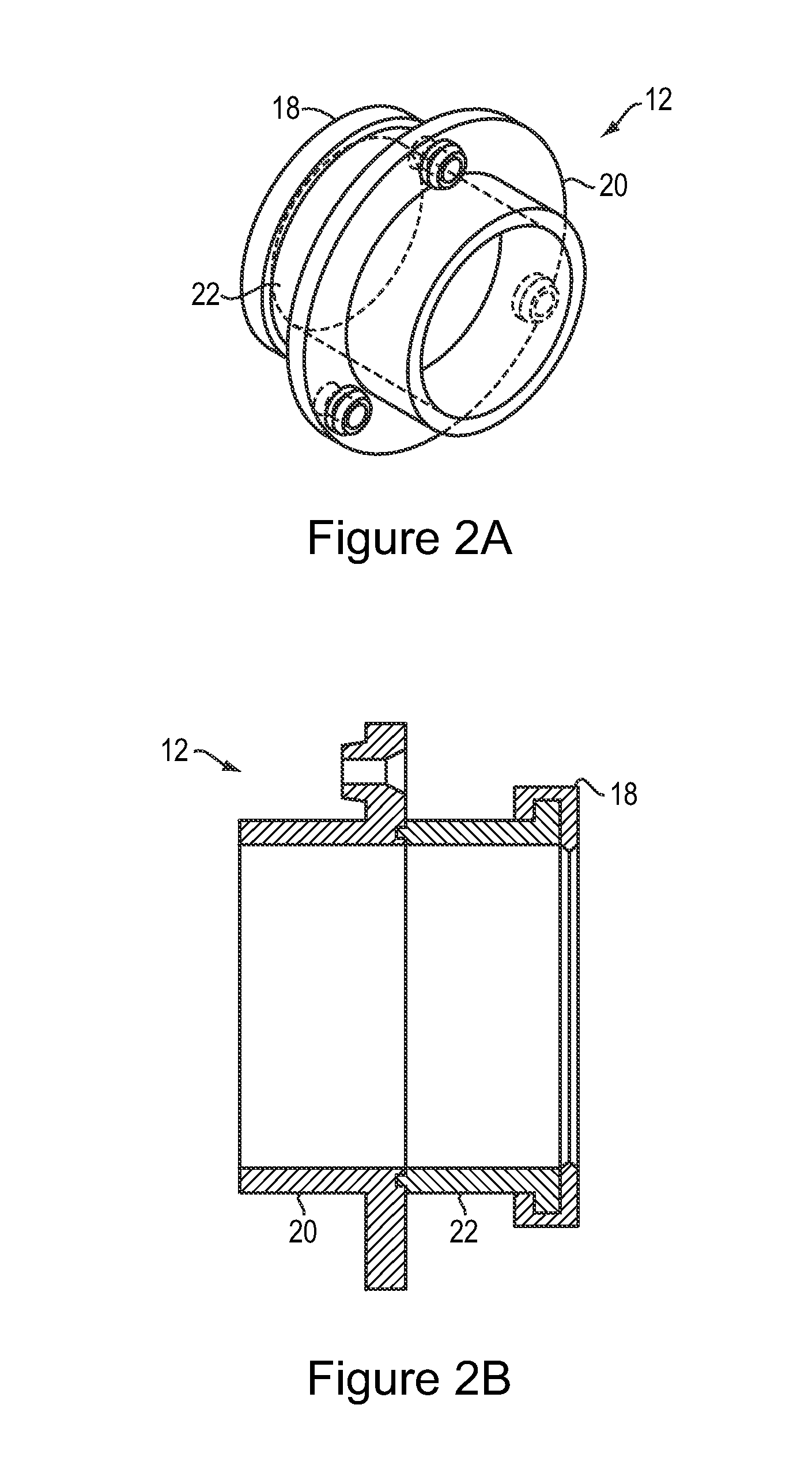
[0026]FIGS. 1A-1C depict a device 10 for providing access to the solid matter (for example, tumor tissue, a blood clot, a cyst, a brain lesion or infected brain tissue) disposed within the brain tissue of a subject. The device 10 includes a cranial anchor 12, a channel member 14, and an optional trocar 16. An anchor seal 18 may optionally be provided as part of the anchor 12. The channel member 14 is dimensioned to fit within an anchor passage of the anchor 12 and to receive the trocar 16. A tight fit between the anchor 12 and the channel member 14 reduces the likelihood that channel member 14 will become misaligned from the anchor 12. The anchor seal 18, when present, can also help stabilize the channel member 14 as it passes through ...


PUM


Abstract

The present invention relates to a method and device for removing solid matter from a brain and controlling bleeding associated with the removal of the matter. The method involves securing a cranial anchor to a region of the skull in which an opening has been created to expose brain matter and introducing through a passage defined by the anchor a channel member that displaces brain tissue and exposes the solid matter. After removing the solid matter, a flowable hemostat is introduced into the cavity created by removal of the matter. A balloon introduced into the working channel is then inflated to compress the hemostat against the wall of the cavity to control bleeding from blood vessels around the cavity. The device includes a cranial anchor, a channel member defining a working channel, an optional removable trocar, and a catheter for introducing the hemostat and the inflatable balloon into the cavity.

Description

CROSS-REFERENCE TO RELATED APPLICATION[0001]This application claims the benefit of and priority to U.S. Provisional Patent Application Ser. No. 61 / 496,803, filed on Jun. 14, 2011, the disclosure of which is incorporated by reference herein in its entirety.FIELD OF THE INVENTION[0002]The invention relates to a method and system for removing solid matter disposed within brain tissue. More particularly, the invention relates to a method and system for facilitating the removal of solid matter from the brain and for controlling bleeding with a flowable hemostat once the solid matter has been removed.BACKGROUND[0003]Occasionally undesirable solid matter develops in brain tissue, and can include, a blood clot (also known as an intracranial hemorrhage), tumor tissue, a cyst, a brain lesion or infected brain tissue. Depending upon the size and location of the solid matter, and the condition of the subject, it may be necessary to remove the solid matter via surgical intervention.[0004]Given t...

Claims


Application Information

Patent Timeline
Patent Type & Authority Patents(United States)
IPC IPC(8): A61F2/958A61M31/00
CPCA61B17/3498A61B2019/208A61B17/3421A61B17/3472A61B2017/3492A61B17/0218A61B17/50A61M25/10A61B2090/103
Inventor ABRAHAMS, JOHN, M.
Owner ABRAHAMS JOHN M
Why Eureka
Try Eureka