Density-based cell detection system

a density-based cell and detection system technology, applied in biochemistry apparatus and processes, instruments, specific gravity measurement, etc., to achieve the effect of low cost and convenien

Active Publication Date: 2014-03-25
BECTON DICKINSON & CO
View PDF12 Cites 0 Cited by
  • Summary
  • Abstract
  • Description
  • Claims
  • Application Information

AI Technical Summary

Benefits of technology

[0004]The invention provides a low-cost and convenient method for detecting certain cell types in a sample by separating them from other cell types using density labels, such as gold particles. In one aspect, the invention provides a method for detecting a subpopulation of cells in a sample, wherein cells of the subpopulation have characteristic numbers of one or more cell surface markers, comprising the following steps: (a) combining under probe-binding conditions a sample with a probe comprising at least one binding compound specific for at least one of the one or more cell surface markers and a particle having a size and a composition, thereby forming a reaction mixture containing labeled cells from the subpopulation and other cells, wherein the one or more cell surface markers are present in greater numbers on cells of the subpopulation than on other cells of the sample and wherein the one or more cell surface markers, and the particle size and composition are selected so that labeled cells of the subpopulation have a density that is greater than that of other cells of the sample; (b) separating labeled cells of the reaction mixture through a density medium, the labeled cells of the subpopulation being denser than the density medium and the other cells of the sample being less dense than the density medium, so that the labeled cells of the subpopulation are separated from the other cells; and (c) detecting the subpopulation of cells in the density medium.
[0005]In another aspect, such method further provides a step of separating the reaction mixture through said density medium disposed in a capillary having a bottom, so that said labeled cells from the subpopulation form a stack having a height from the bottom of the capillary, wherein the height of the stack is monotonically related to the number of cells of the subpopulation in said sample. An exemplary, density medium is Histodenz™, or like media. Preferably, in such aspect, the stack of cell can be seen with the unaided eye. In further preference, the density medium is colored with a contrasting color to that of the density label, so that visualization of the stack is enhanced whenever the stack of cells displaces the density medium at the bottom of the capillary.

Problems solved by technology

Although such advances have reduced costs of cell counting techniques, generally all of these approaches still require detection or sample handling systems that either have dedicated electronic or optical components needing trained operators, calibration, or external power, or other drawbacks, or still do not reduce costs to levels permitting widespread applicability.

Method used


Image

  • Density-based cell detection system
  • Density-based cell detection system
  • Density-based cell detection system

Examples


Embodiment Construction

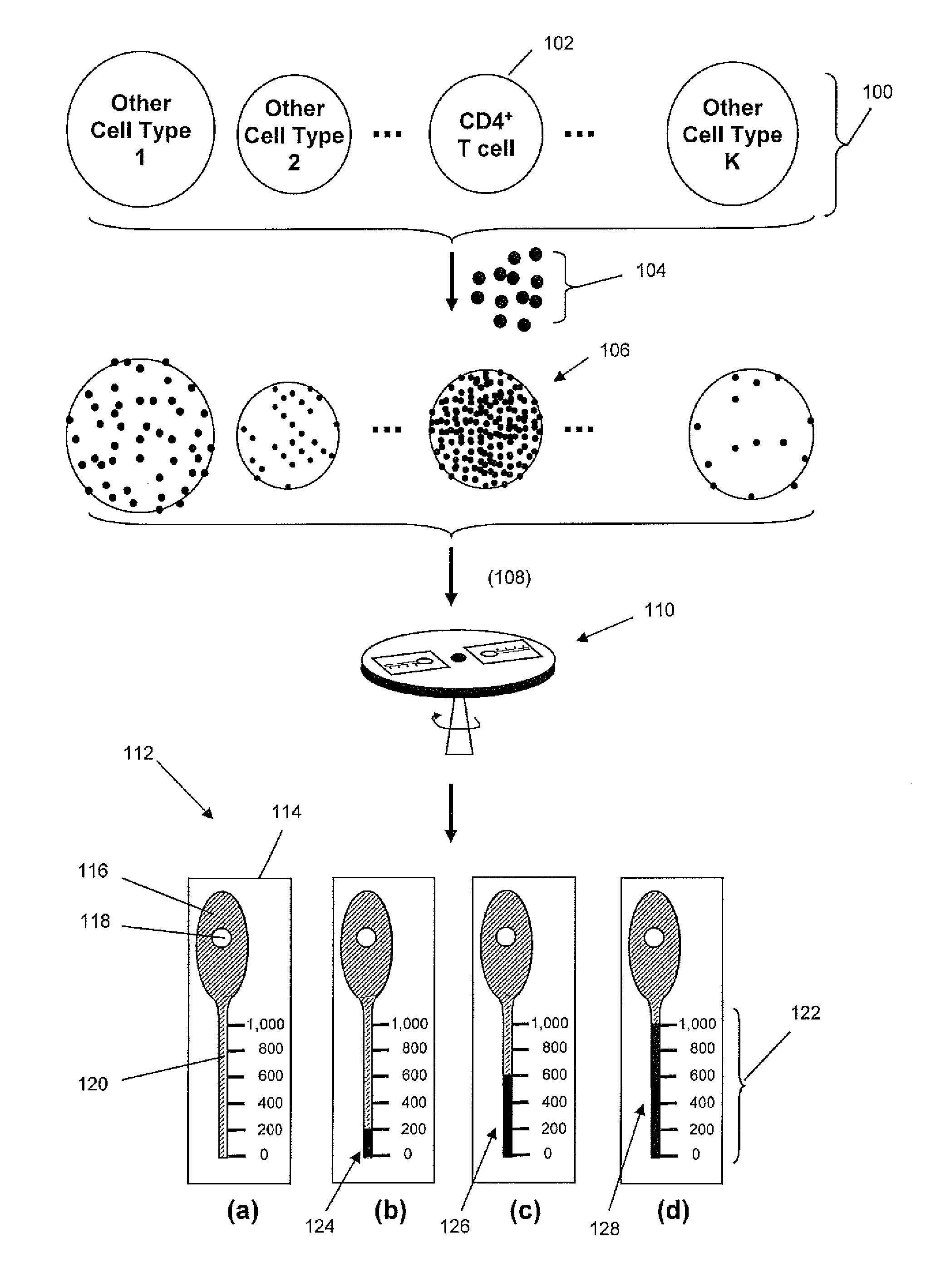
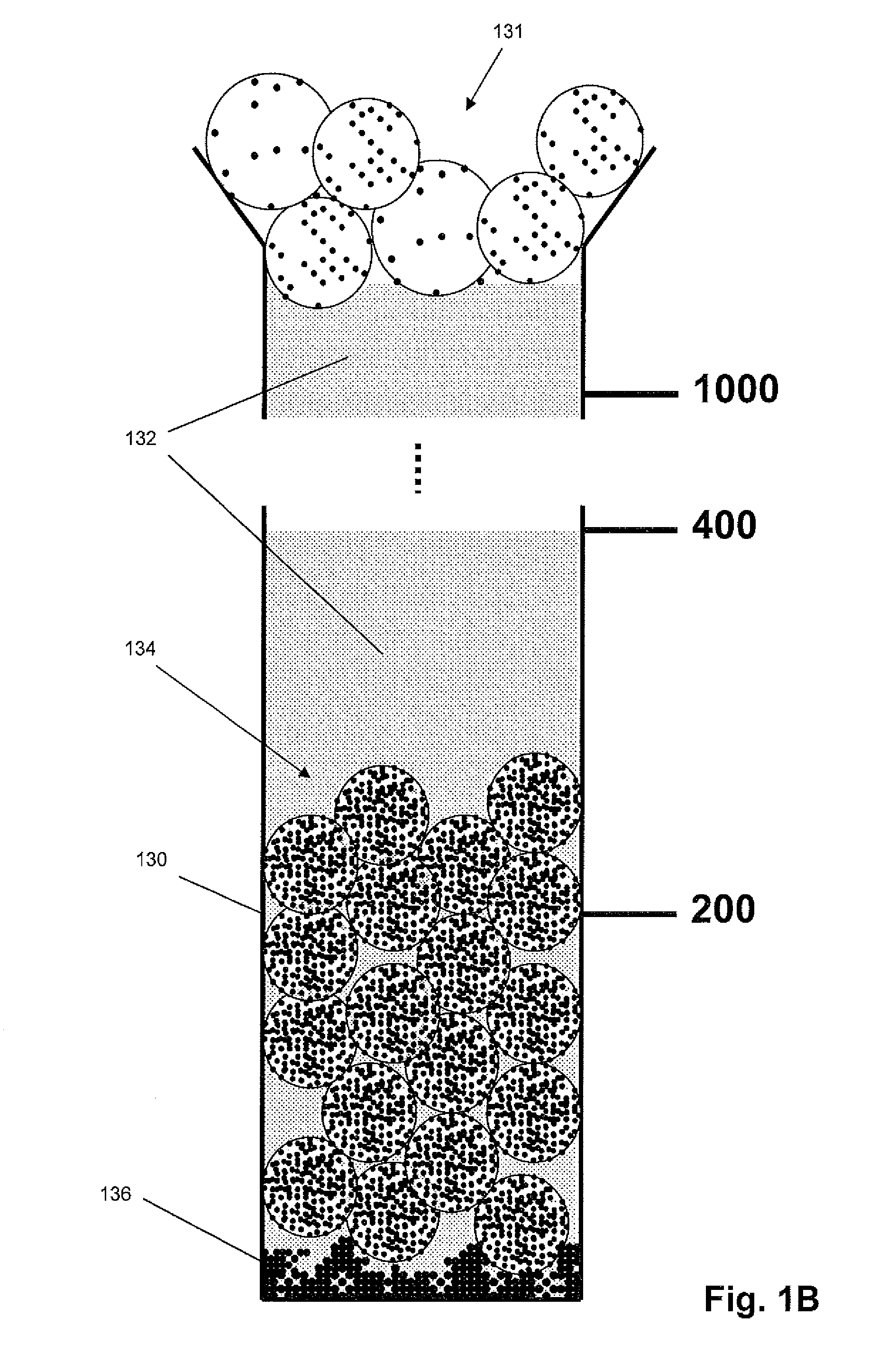
[0012]The practice of the present invention may employ, unless otherwise indicated, conventional techniques from molecular biology (including recombinant techniques), cell biology, immunoassay technology, microscopy, and analytical chemistry, which are within the skill of the art. Such conventional techniques include, but are not limited to, detection of fluorescent signals, image analysis, selection of illumination sources and optical signal detection components, selecting antibodies for specific cellular or molecular targets; labeling of biological cells, and the like. Such conventional techniques and descriptions can be found in standard laboratory manuals such as Genome Analysis: A Laboratory Manual Series (Vols. I-IV), Using Antibodies: A Laboratory Manual, Cells: A Laboratory Manual, PCR Primer: A Laboratory Manual, and Molecular Cloning: A Laboratory Manual (all from Cold Spring Harbor Laboratory Press); Wild, Editor, The Immunoassay Handbook, Third Edition (Elsevier Science,...


PUM

PropertyMeasurementUnit
sizeaaaaaaaaaa
sizesaaaaaaaaaa
sizesaaaaaaaaaa

Abstract

The invention provides a low-cost method for identifying and / or measuring a subpopulation of cells in a biological sample, such as a subpopulation of CD4-positive cells in a whole blood sample. In accordance with the invention, one or more cell surface markers are identified on a subpopulation of interest, which collectively are present in higher numbers on the cells of the subpopulation than on cells not in the subpopulation of interest. A probe comprising a binding compound and a density label are combined with the sample and the resulting mixture is centrifuged or sedimented through a density medium in a capillary. Labeled cells of interest pass through the density medium and stack at the bottom of the capillary where they may be visually detected and quantified by the height of the stack.

Description

CROSS-REFERENCE[0001]This application claims the benefit of U.S. Provisional Application No. 61 / 069,078, filed Mar. 11, 2008, which application is incorporated herein by reference.BACKGROUND[0002]The majority of HIV-infected people live in resource-poor regions where instruments critical for detecting, staging, and monitoring the disease condition resulting from HIV are frequently unavailable because of difficult field conditions, cost, or lack of trained personnel, e.g. Cohen, AIDS, Suppl 4: S81-S87 (2007). This has led to the development by several groups of low-cost instrumentation designed for deployment in resource-poor settings for measuring CD4 counts in patient blood samples, e.g. Rodriguez et al, PLoS Medicine, 2(7): e182 (2005); Ymeti et al, Cytometry A 71: 132-134 (2007); Cheng et al, Lab Chip, 7(2): 170-178 (2007); Pattanapanyasat et al, Clinical Cytometry, 72B: 387-396 (2007); and the like. Although such advances have reduced costs of cell counting techniques, generally...

Claims


Application Information

Patent Timeline
Patent Type & Authority Patents(United States)
IPC IPC(8): G01N33/50G01N15/04G01N9/36
CPCG01N33/5375G01N33/54313C12Q2563/155G01N15/042G01N33/56972G01N9/36G01N2333/70514
Inventor KIRAKOSSIAN, HRAIR
Owner BECTON DICKINSON & CO
Why Eureka
Try Eureka