

Markers and diagnostic methods for metastasis
a technology of metastasis and diagnostic methods, applied in the field of prediction, prognosis or diagnosis of metastasis, can solve the problem of not achieving enough clinical significance, and achieve the effect of higher expression
- Summary
- Abstract
- Description
- Claims
- Application Information
AI Technical Summary
Benefits of technology
Problems solved by technology
Method used
Image

Examples
example 1
Determine of Differentially Expressed Protein Profiles in Lymph Node Positive Vs Lymph Node Negative Breast Cancers
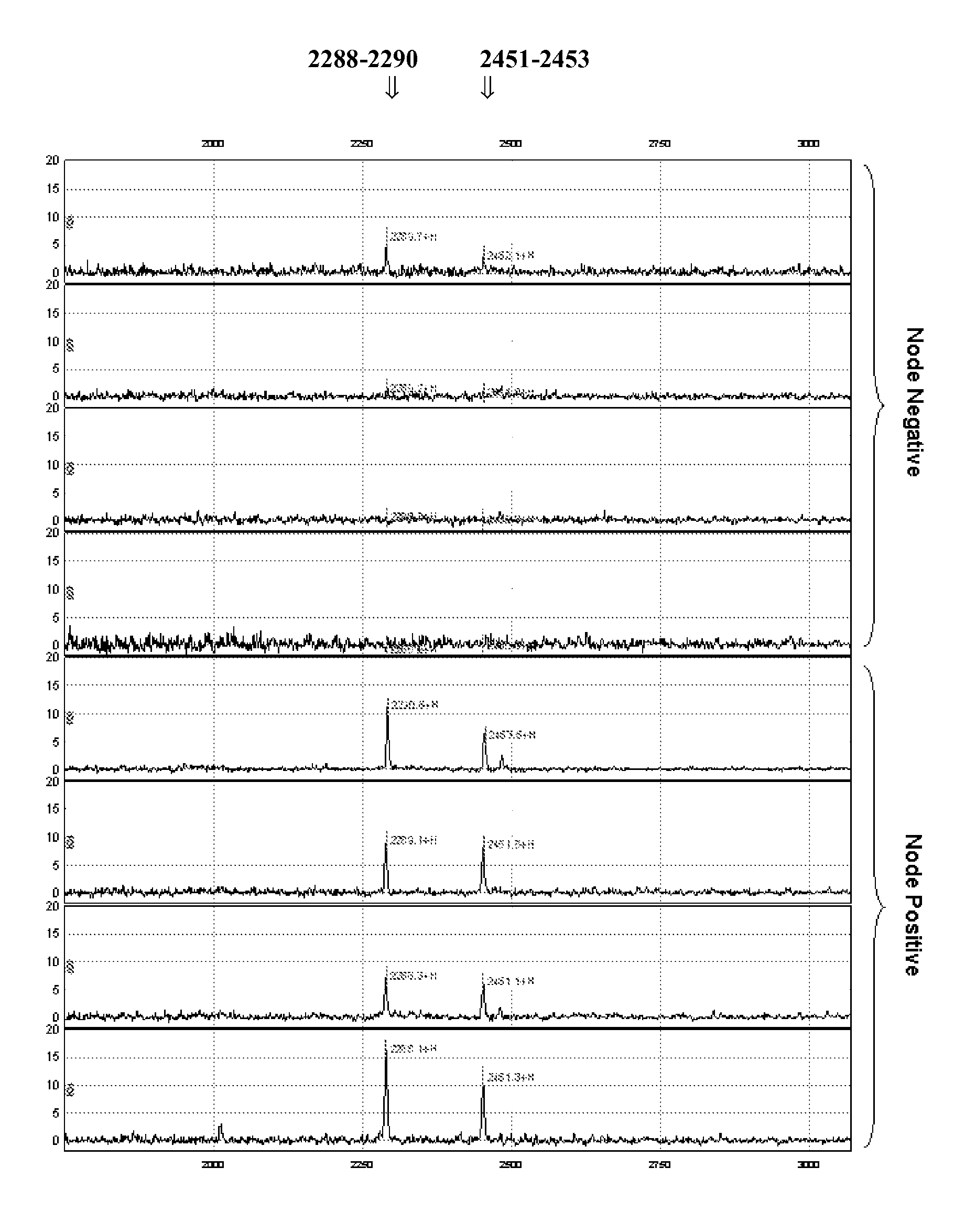
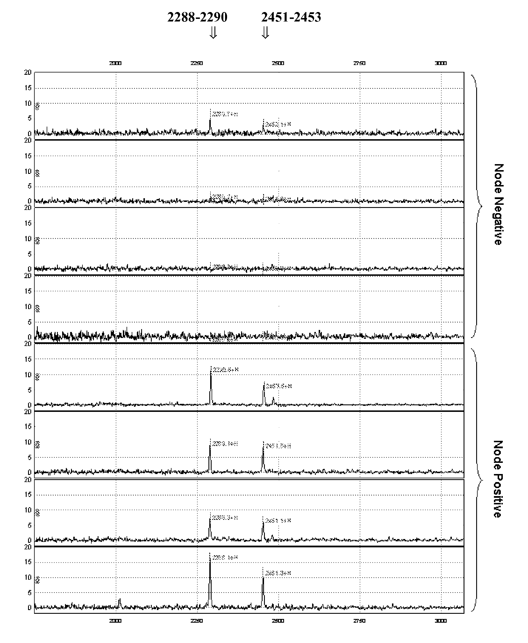
[0108]Because lymph node involvement is often the earliest sign of tumour progression, insights into the underlying molecular mechanisms are essential in order to be able to predict lymph node involvement from protein expression profiles, more in particular of primary breast tumour tissue. A pilot proteomic study in 8 patients with breast cancer was performed, investigating the differences in protein expression profiles between lymph node negative and lymph node positive breast cancers using SELDI-TOF MS. Representative biopsies were selected taking various clinicopathological and biological parameters into account.
[0109]Surface Enhanced Laser Desorption / Ionization Time-of-flight Mass Spectrometry (SELDI-TOF MS) is a valuable method for the analysis of differential protein expression in tissue extracts and body fluids (Bischoff R. et al. 2004 J. Chromatogr. B Analyt. Te...
example 2
Identification of Differentially Expressed Proteins
[0122]For the identification of the proteins listed in Table 2, standard methods and protocols known in the art can be used. The different proteins can be collected individually and than sequenced with an automated amino acid sequencer.
Materials and Methods
Peptide Purification
[0123]250 μl of tissue extract was diluted with 250 μl 50 mM Tris-HCl pH 9 and loaded on an anion exchange mini spin column (Sartorius, Aubange, France) that was equilibrated with 500 μl 50 mM Tris-HCl pH 9. After binding of the peptides to the membrane, the column was washed three times with 500 μl 50 mM Tris-HCl pH 9 and the peptides were finally eluted with 500 μl 50% Acetonitrile / 1% Formic Acid. The eluate was subsequently evaporated in a vacuum centrifuge and re-dissolved in 10 μl 500 mM Tris-HCl pH 9.
[0124]5 μl of this purified and concentrated fraction of anionic peptides was then loaded on an anion exchange ProteinChip array and incubated in a humid cha...
example 3
Two-Dimensional Gel Electrophoresis Followed by Protein Identification
Materials and Methods
[0135]All materials, reagents and software for 2 Dimensional Differential Gel Electrophoresis (2D DIGE) were purchased from GE Healthcare (London, UK).
Sample Preparation for 2D DIGE Analysis
[0136]In this study, 12 breast carcinoma tissue extracts were used, 6 lymph node metastasis positive and 6 lymph node metastasis negative control samples. 50 μg of each tissue protein extract was labeled with 200 pmol of Cy3 or Cy5 (two “forward” and two “reverse” labeled samples were prepared for the two sample groups), whereas 50 μg of pooled internal standard was labeled with 200 pmol of Cy2. The internal standard consisted of a pool of all lymph node positive and negative breast carcinoma tissue extracts. The labeling reaction was carried out for 30 min on ice and quenched with 10 mM lysine (15 min on ice). Labeled protein extracts were pooled, and sample loading buffer was added (7 M urea, 2 M thiourea...
PUM
 Abstract
Description
Claims
Application Information