Device, system and method for in vivo analysis

a technology devices, applied in the field of in vivo analysis, can solve the problems of inability to detect early, limited detection, and pathologies in other parts of the gi tract, such as, for example, the small intestine, which may not be easily detected by endoscopy

Active Publication Date: 2014-05-27
GIVEN IMAGING LTD
View PDF123 Cites 8 Cited by
  • Summary
  • Abstract
  • Description
  • Claims
  • Application Information

AI Technical Summary

Problems solved by technology

One of the drawbacks of this method is that the appearance of antibodies in the blood stream may usually occur at a late stage of the disease, such that early detection may not be possible.
Detection of some pathologies in the GI tract may be performed using an endoscope; however, such detection may be limited to the upper or lower gastrointestinal tract.
Thus, pathologies in other parts of the GI tract, such as, for example, the small intestine, may not be easily detected by endoscopy.
Typically these in vitro methods of detection are carried out in a lab using cumbersome and complicated machines.
Complicated machines may be needed, for example, because of the complicated requirements of such procedures which may involve multiple reactions and / or different conditions for each reaction, and typically washing in between reactions.
Furthermore, these methods of detection, when carried out in vitro, may not easily enable the localization or identification of the origin of an abnormally occurring substance.

Method used


Image

  • Device, system and method for in vivo analysis
  • Device, system and method for in vivo analysis
  • Device, system and method for in vivo analysis

Examples


Embodiment Construction

[0053]In the following detailed description, numerous specific details are set forth in order to provide a thorough understanding of the invention. However, it will be understood by those skilled in the art that the present invention may be practiced without these specific details. In other instances, well-known methods, procedures, and components have not been described in detail so as not to obscure the present invention.

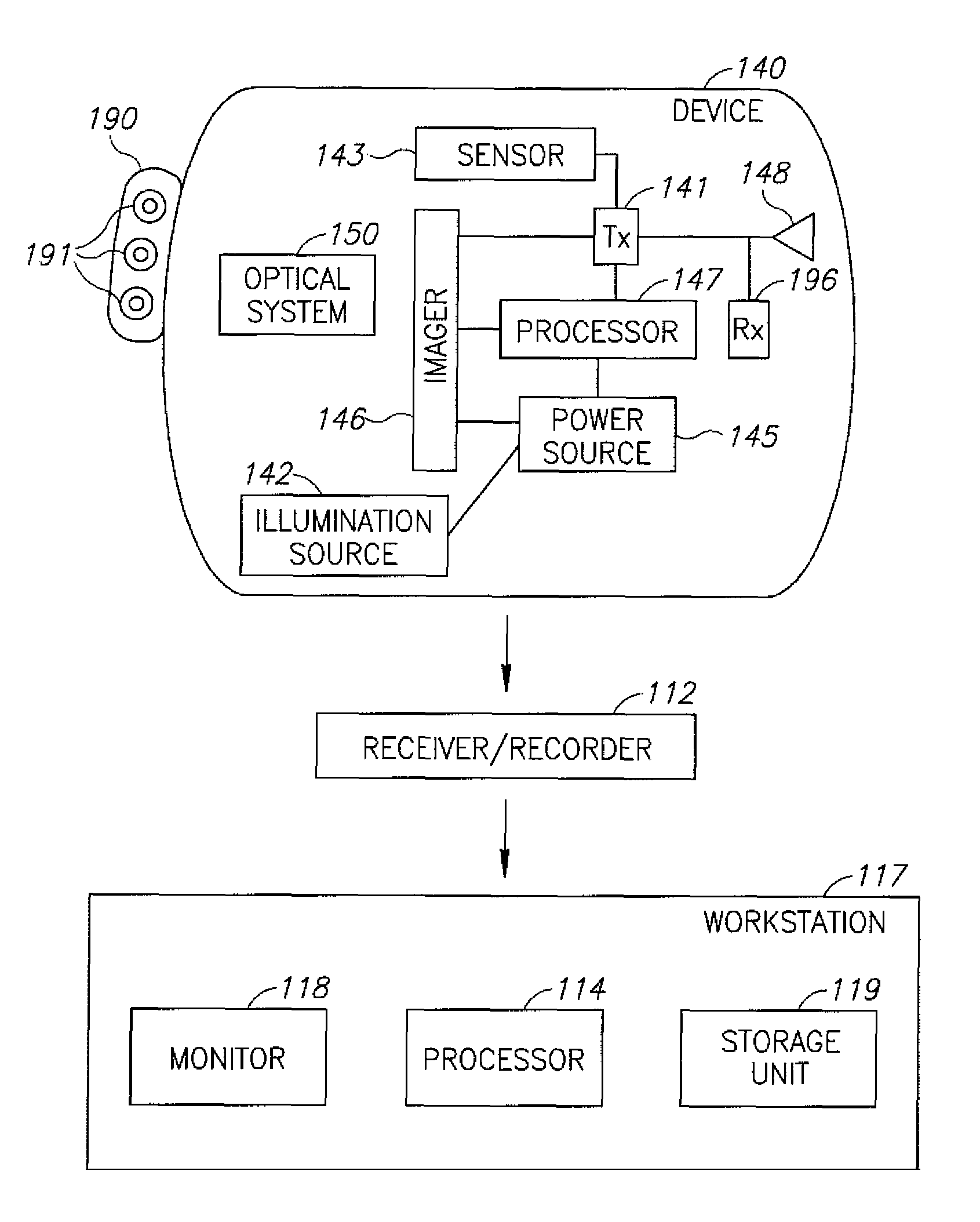
[0054]Some embodiments of the present invention are directed to a typically one time use or partially single use detecting device, which may be used as an in vitro analysis kit. Other embodiments of the present invention are directed to a typically in vivo device, e.g. a swallowable device that may passively or actively progress through the gastro-intestinal (GI) tract, pushed along, in some embodiments, by natural peristalsis. Some embodiments are directed to in vivo sensing devices that may be passed through other body lumens such as, for example, through blood ...


PUM


Abstract

A device for in vivo analysis includes: a reaction chamber to store a detecting reagent able to react with a sample collected in vivo; and optionally a labeled-substance chamber to store a labeled substance able to bind to at least a portion of a compound resulting from a reaction of the detecting reagent and the sample. The in-vivo imaging device, typically an autonomous capsule, may have a housing, the housing comprising a window; an illumination source located within the housing to illuminate a body lumen through the window; an imager to receive light reflected from the body lumen through the window; and a transmitter to transmit image data to a receiving system. The window is coated with liposomes containing a marker such that the imager may acquire images which include the marking.

Description

CROSS-REFERENCE TO RELATED APPLICATIONS[0001]This application is a Continuation-in-Part under 35 U.S.C. §120 of PCT International Application No. PCT / IL2006 / 000127, International Filing Date Jan. 31, 2006, which claimed priority from U.S. Patent Application No. 60 / 647,790, filed Jan. 31, 2005. This application is also a Continuation-in-Part under 35 U.S.C. §120 of PCT International Application No. PCT / IL2007 / 000458, International Filing Date Apr. 10, 2007, which claimed priority from U.S. Patent Application No. 60 / 788,114, filed Apr. 3, 2006 which is hereby incorporated by reference.FIELD OF THE INVENTION[0002]The present invention relates in general to in vivo analysis, and specifically to in vivo analysis utilizing micro-fluidic techniques of body fluids possibly on a solid state substrate and utilizing liposomes and / or nano-containers.BACKGROUND OF THE INVENTION[0003]An atypical concentration or presence of substances in body fluids or in body lumens may be indicative of the biol...

Claims


Application Information

Patent Timeline
Patent Type & Authority Patents(United States)
IPC IPC(8): A61B5/1455
CPCA61K49/0084A61B5/073A61K49/0093A61K49/0002A61B1/041
Inventor RABINOWITZ, ELISHAKATZ, EMIL-ISRAEL
Owner GIVEN IMAGING LTD
Why Eureka
Try Eureka