Aberration correction using channel data in ultrasound imaging system

a technology of ultrasound imaging system and channel data, applied in the field of ultrasound imaging system, can solve the problems of limited space available on the console for imaging control keys, difficult to use, and difficult to design the user-control system

Active Publication Date: 2014-07-22
SHENZHEN MINDRAY BIO MEDICAL ELECTRONICS CO LTD
View PDF75 Cites 43 Cited by
  • Summary
  • Abstract
  • Description
  • Claims
  • Application Information

AI Technical Summary

Benefits of technology

[0017]In accordance with another aspect of the invention, an ultrasound system comprises a channel data memory to store channel data obtained by digitizing ultrasound echo data produced by an image scan; an image data processor configured to process the stored channel data in the memory to reconstruct an ultrasound image for each of a plurality of trial values of at least one parameter to be optimized; and a parameter optimization unit configured to evaluate an image quality of the reconstructed ultrasound image for each trial value of the at least one parameter, and to determine the optimized value of the at least one parameter based on the evaluated image quality.
[0018]In accordance with another aspect of the present invention, an ultrasound system comprises a channel data memory to store channel data obtained by digitizing ultrasound echo data produced by an image scan; an image data processor configured to process the stored channel data in the memory to reconstruct an ultrasound image for each of a plurality of trial values of at least one parameter to be optimized; and a parameter optimization unit including a parameter optimization program stored in a computer readable storage medium. The parameter optimization program includes code for evaluating an image quality of the reconstructed ultrasound image for each trial value of the at least one parameter, and code for determining the optimized value of the at least one parameter based on the evaluated image quality.

Problems solved by technology

The need to optimize both kinds of system parameters is a long-standing challenge in diagnostic ultrasound imaging, mainly because (1) the majority of sonographers or users often lack the time and / or training to properly operate a very broad range of user-controls for optimal system performance; and (2) engineering presets are usually pre-determined by the manufacturer based on “typical” or “average” system operating conditions including patient type (body size and fat / muscle composition), normal and abnormal tissue characteristics for various application types, and environmental factors (e.g., ambient light condition).
For a compact scanner, user-control design is particularly challenging because the space available on the console for imaging control keys can be very limited.
This means that the overall user-control subsystem will be restricted and / or more difficult to use (e.g., accessing multiple layers of soft-key menus) compared to conventional cart-based scanners.
Another related challenge for all ultrasound scanners is ergonomics.

Method used


Image

  • Aberration correction using channel data in ultrasound imaging system
  • Aberration correction using channel data in ultrasound imaging system
  • Aberration correction using channel data in ultrasound imaging system

Examples


Embodiment Construction

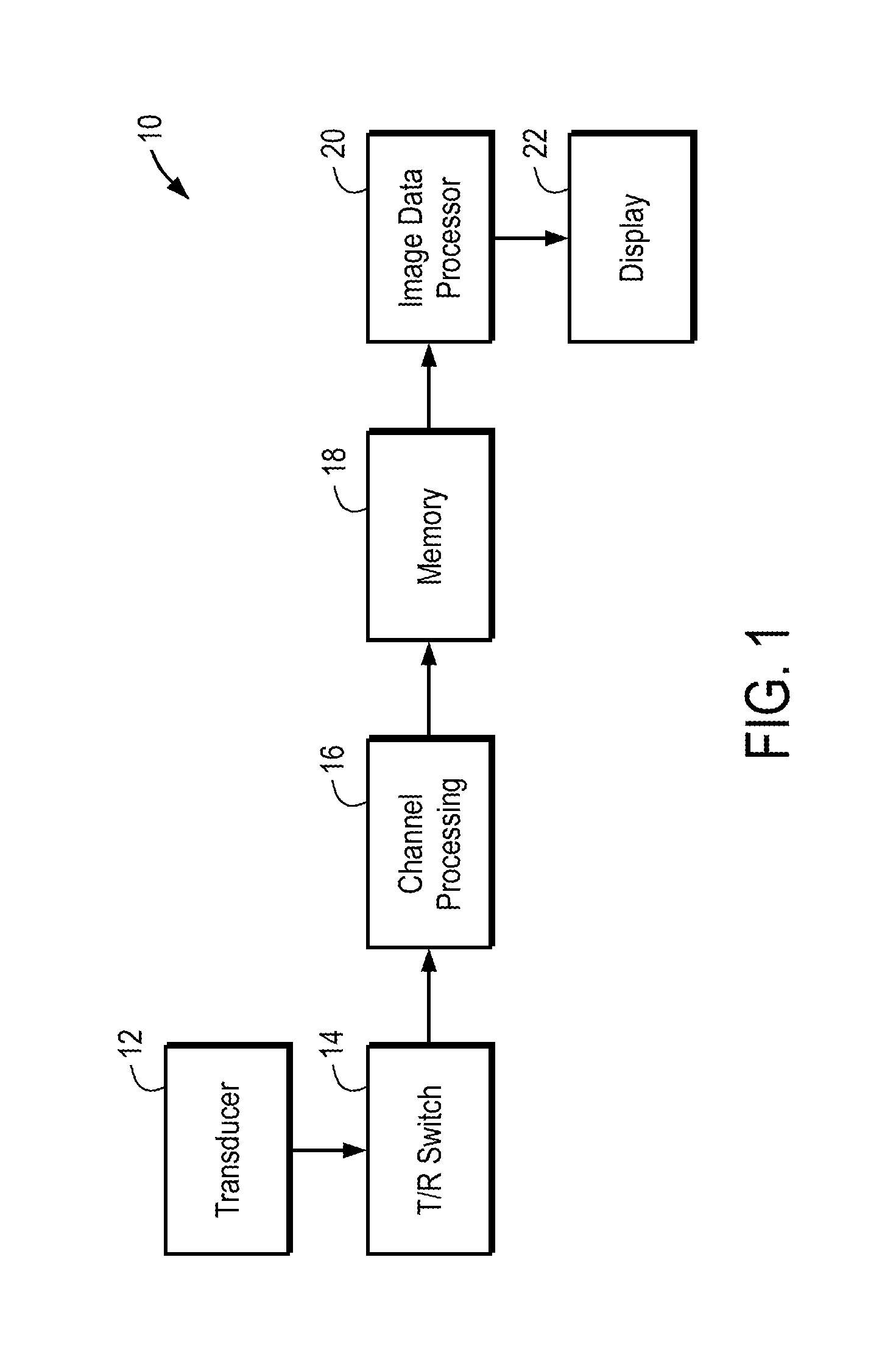
[0027]FIG. 1 is a block diagram of the system architecture of an ultrasound scanner 10. In a specific embodiment, the scanner 10 employs a zone-based technique for real-time B-mode imaging, and utilizes a transmit beam from which many receive beams are formed, such that a full field of view image can be formed using much fewer firings than a conventional system, i.e., 30-50 firings. The main signal path begins with a transducer 12 with switch 14. The transducer 12 includes an array of transducer elements. For each transmit beam firing, the receive RF echo data is digitized and preprocessed within the channel processing block 16 where demodulation, averaging, and filtering occurs. Next, the baseband channel in-phase / quadrature (I / Q) data is stored in a large memory 18. An image data processor 20 (e.g., one or more DSPs or Digital Signal Processors) pulls the channel I / Q data from the memory 18, and performs dynamic receive beam formation, detection, log compression, filtering, and sc...


PUM

PropertyMeasurementUnit
sound speedaaaaaaaaaa
speedaaaaaaaaaa

Abstract

Embodiments of the present invention provide an ultrasound scanner equipped with an image data processing unit that can perform adaptive parameter optimization during image formation and processing. In one embodiment, an ultrasound system comprises a channel data memory to store channel data obtained by digitizing ultrasound image data produced by an image scan; an image data processor configured to process the stored channel data in the memory to reconstruct an ultrasound image for each of a plurality of trial values of at least one parameter to be optimized; and a parameter optimization unit configured to evaluate an image quality of the reconstructed ultrasound image for each trial value of the at least one parameter, and to determine the optimized value of the at least one parameter based on the evaluated image quality.

Description

CROSS-REFERENCES TO RELATED APPLICATIONS[0001]This application claims the benefit of U.S. Provisional Patent Application No. 60 / 701,635, filed Jul. 22, 2005, the entire disclosure of which is incorporated herein by reference.BACKGROUND OF THE INVENTION[0002]This invention relates generally to ultrasound imaging systems and, more particularly, to adaptive optimization of ultrasound imaging system parameters by making use of stored channel data.[0003]Medical ultrasound imaging systems need to support a set of imaging modes for clinical diagnosis. The basic imaging modes are timeline Doppler, color flow velocity and power mode, B-mode, and M-mode. In B-mode, the ultrasound imaging system creates two-dimensional images of tissue in which the brightness of a pixel is based on the intensity of the return echo. In color flow imaging mode, the movement of fluid (e.g., blood) or tissue can be imaged. Measurement of blood flow in the heart and vessels using the Doppler effect is well known. T...

Claims


Application Information

Patent Timeline
Patent Type & Authority Patents(United States)
IPC IPC(8): G01N29/44G01S7/52A61B8/00G06T5/50A61B8/08
CPCG01S7/52049A61B8/5269A61B8/58G01N29/4463A61B8/00G01S7/52085G01S7/52095G06T5/50G06T2207/10132G06T2207/20172G06T2207/30004A61B8/145G01S7/52046A61B8/585A61B8/488A61B8/469A61B8/467A61B8/5207A61B8/4483G06T7/0022G06T11/005A61B8/461G06T7/97
Inventor NAPOLITANO, DAVID J.CHOU, CHING-HUAJI, TING-LANDEBUSSCHERE, BRIAN DEREKMCLAUGHLIN, GLEN W.MO, LARRY Y. L.STEINS, ROBERT W.
Owner SHENZHEN MINDRAY BIO MEDICAL ELECTRONICS CO LTD
Why Eureka
Try Eureka