Method and apparatus for protection from high intensity light

a technology of high intensity light and protection apparatus, applied in the direction of optical radiation measurement, instruments, television systems, etc., can solve the problems of affecting the image quality of the image, affecting the image quality, and affecting the safety of patients, so as to minimize the number of increments and optimize the effect of image quality

Active Publication Date: 2014-11-04
KARL STORZ IMAGING INC
View PDF45 Cites 7 Cited by
  • Summary
  • Abstract
  • Description
  • Claims
  • Application Information

AI Technical Summary

Benefits of technology

[0017]Accordingly, it is an object of the present invention to provide a method and apparatus to overcome known problems when working to control light sources that have slow-response, high-frequency noise, nonlinearity, and non-monotonic response times.
[0018]It is an object of the invention to provide a method for upgrading existing and newly-designed endoscopic imaging systems with a light source control (“LSC”) feature that controls the intensity of the light source output. It is an object of the invention to reduce and / or eliminate costs associated with upgrading of existing and newly-designed endoscopic imaging systems with the LSC feature without any hardware change in product lines of light sources, camera-heads / imagers, CCUs, videoscopes, and endoscopes.

Problems solved by technology

The use of high intensity light sources involves potential hazards to medical personnel and patients.
For example, when a light guide cable, used to convey the high intensity-light source output, is momentarily disconnected from the endoscope and placed on a sterile drape used to protect the patient, the high intensity of light output can be sufficient to ignite the drape and pose a fire hazard; or, the user can inadvertently hold the disconnected light guide cable in such a way as to temporarily blind another person in the room.
In some instances, when the endoscope is removed from (i.e., pulled out of) a patient, there can be a risk of these same hazards.
This increased light intensity level can desiccate body tissue and cause serious injury to the patient.
An undesirable, and potentially dangerous, scenario can occur if the light source output is set to a high intensity level, and the endoscope distal end is placed within close proximity to tissue being imaged.
In such a situation, the risk of desiccating delicate tissue is greatly increased.
These safety solutions are not necessarily effective against all potential hazardous conditions that may arise; such as when the endoscope with the light guide cable still attached is pulled out of a patient and inadvertently directed at a person or surgical drape, or when the light guide or source initially is directed to treat openly accessible tissue and inadvertently misdirected during or after a procedure, or when a video camera head, attached to the endoscope light guide cable combination, is disconnected from its corresponding control unit.
However, Chatenever '422, while effective as a safety solution, has problems controlling Xenon lights and other light sources because the amplitude or frequency modulation methods described by Chatenever '422 do not work well with light sources having these problems.
Specifically, Chatenever '422 does not work well with lights sources that have slow-response, high-frequency noise, nonlinearity, and non-monotonic response times, such as Xenon lights.
None of the other existing methods and apparatuses described in the prior art work effectively with light sources that have slow-response, high-frequency noise, nonlinearity, and non-monotonic response times.

Method used


Image

  • Method and apparatus for protection from high intensity light
  • Method and apparatus for protection from high intensity light
  • Method and apparatus for protection from high intensity light

Examples


Embodiment Construction

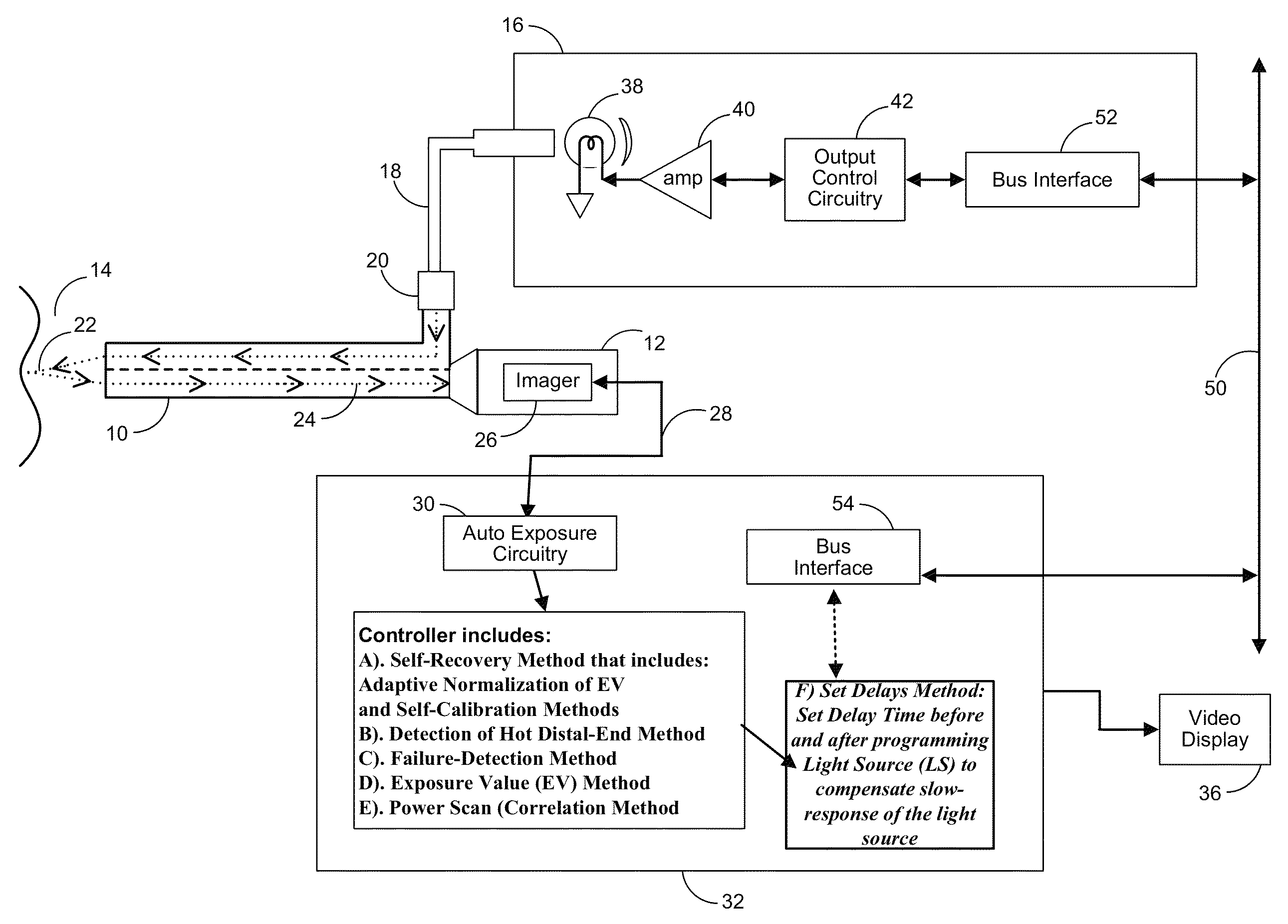
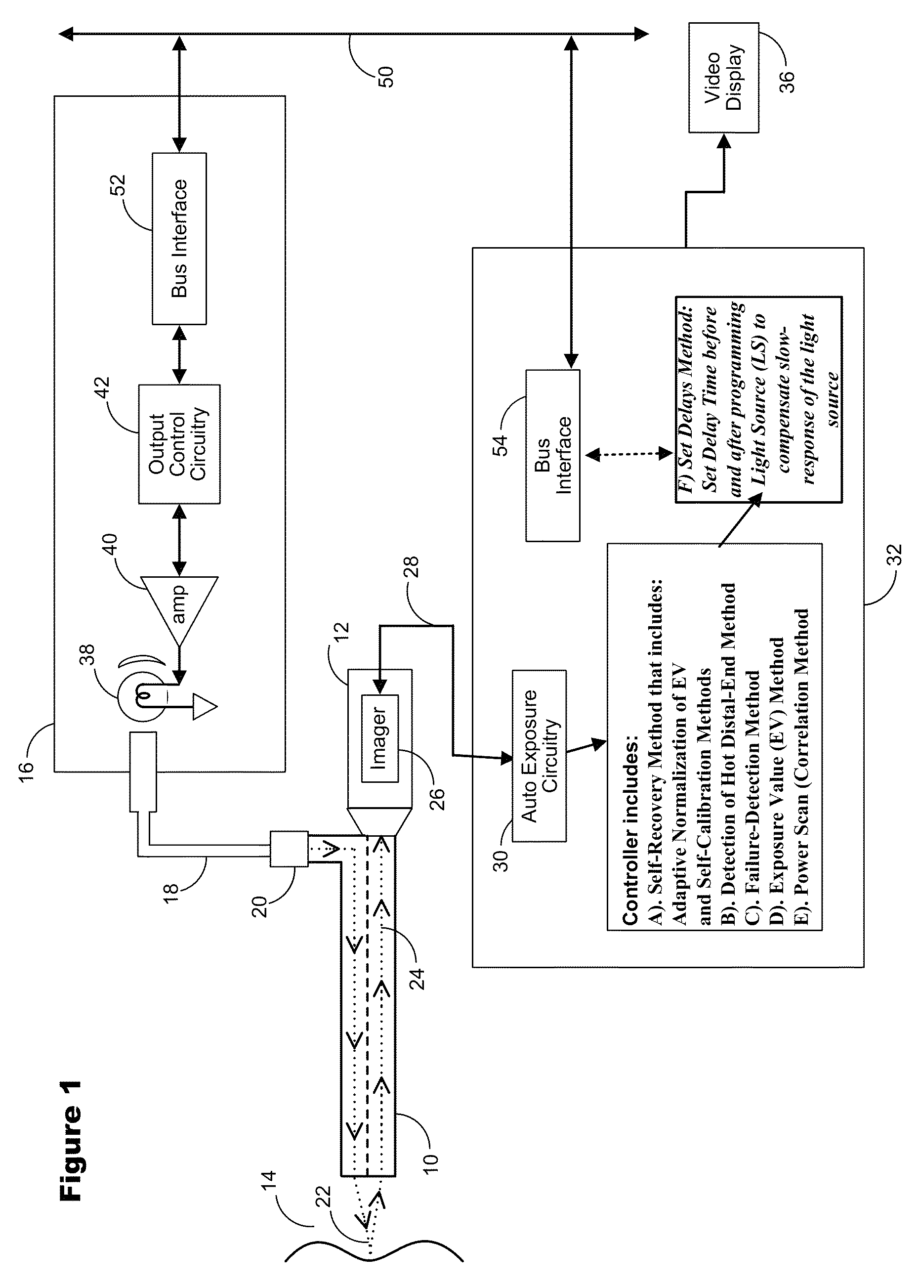
[0097]With reference to FIG. 1, an endoscope 10 is illustrated having a camera head 12 mounted thereto at the proximal end to produce video images in a manner for example as described in the aforementioned U.S. Pat. No. 5,162,913 to Chatenever, et al. The distal end of endoscope 10 is directed at tissue 14 to inspect the tissue with light from a high intensity light source 16 and passed to the distal end through a light guide cable 18. Typically, light guide cable 18 can be disconnected from endoscope 10 at connector 20, thus, posing a safety hazard as described above.

[0098]The light from light guide cable 20 is directed to illuminate tissue 14 as suggested with path 22 and light reflected by tissue 14 is passed along optical path 24 to imager 26 within camera head 12. Imager 26 detects light reflected off tissue 14 by means of optical path 24. Imager 26 may be any type commonly used within the art, such as but not limited to CCD, CID or CMOS imagers. Camera head 12 produces image s...


PUM


Abstract

A method and apparatus where the output from a high intensity light source is controlled to produce well-exposed images / videos and to reduce automatically the intensity when an unsafe issue is detected in medical devices such as endoscopes and the like. The method and apparatus overcome problems to control light sources that have high-frequency noise, slow-response time, nonlinearity, and non-monotonic response time and to protect the patients' tissues from possible overheating / burning and the eyes of personnel and patients from possible direct exposure to high intensity light used in medical devices such as endoscopes and the like.

Description

FIELD OF THE INVENTION[0001]This invention generally relates to a method and apparatus for automatically protecting personnel and patients from direct exposure to the output of a high intensity light source used in medical devices, such as endoscopic imaging systems and the like. The invention also generally relates to a method and apparatus for overcoming problems to control high intensity light sources that have high-frequency noise, slow-response time, nonlinearity, and non-monotonic response time, such as Xenon or Xenon-like based light sources.BACKGROUND OF THE INVENTION[0002]The imaging of body surfaces through an endoscope is well known within the medical and veterinarian fields. Typically, this involves inserting an endoscope into a body cavity and directing a high intensity light source output through the endoscope to illuminate body tissue. Light reflected by the body tissue then is guided along an optical path to an image sensor to generate both video and still images of ...

Claims


Application Information

Patent Timeline
Patent Type & Authority Patents(United States)
IPC IPC(8): A61B1/04G03B15/03A61B1/00H04N5/235G03B15/14H04N7/18A61B1/06
CPCA61B1/042G03B15/03H04N5/2354A61B1/00006A61B1/0669H04N7/18G03B15/14A61B1/06A61B1/00009H04N5/2352A61B1/04A61B1/0655H04N23/72H04N23/74
Inventor OVOD, VLADIMIR, I.
Owner KARL STORZ IMAGING INC
Why Eureka
Try Eureka