Methods and systems for determining a transformation function to automatically register different modality medical images

a technology of transformation function and medical image, applied in the field of medical imaging system, can solve the problems of requiring a significant amount of time and/or computing resources for registration and segmentation operations

Active Publication Date: 2015-09-08
GENERAL ELECTRIC CO
View PDF8 Cites 3 Cited by
  • Summary
  • Abstract
  • Description
  • Claims
  • Application Information

AI Technical Summary

Problems solved by technology

However, the registration and segmentation operations can require a significant amount of time and / or computing resources.

Method used


Image

  • Methods and systems for determining a transformation function to automatically register different modality medical images
  • Methods and systems for determining a transformation function to automatically register different modality medical images
  • Methods and systems for determining a transformation function to automatically register different modality medical images

Examples


Embodiment Construction

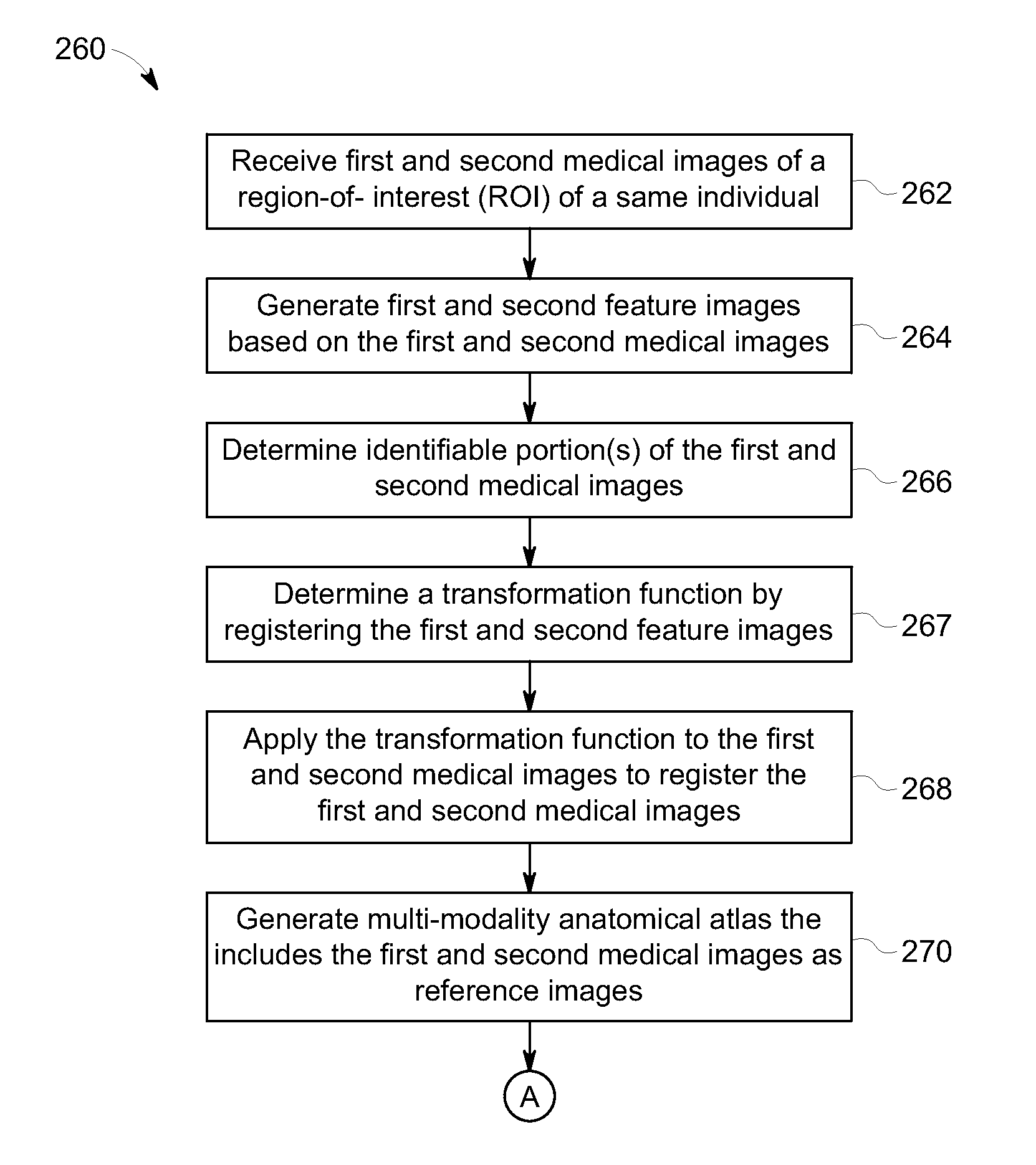
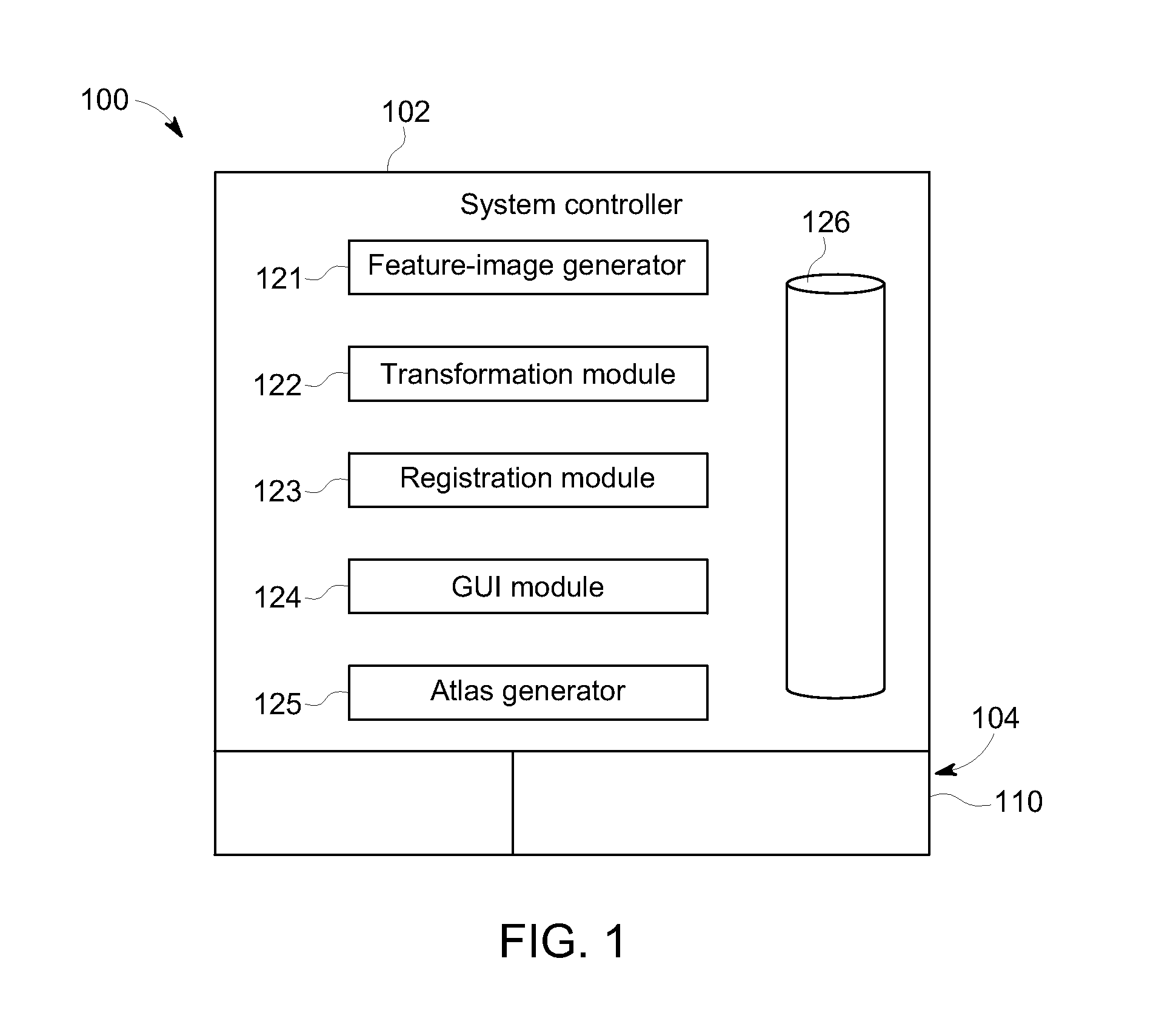
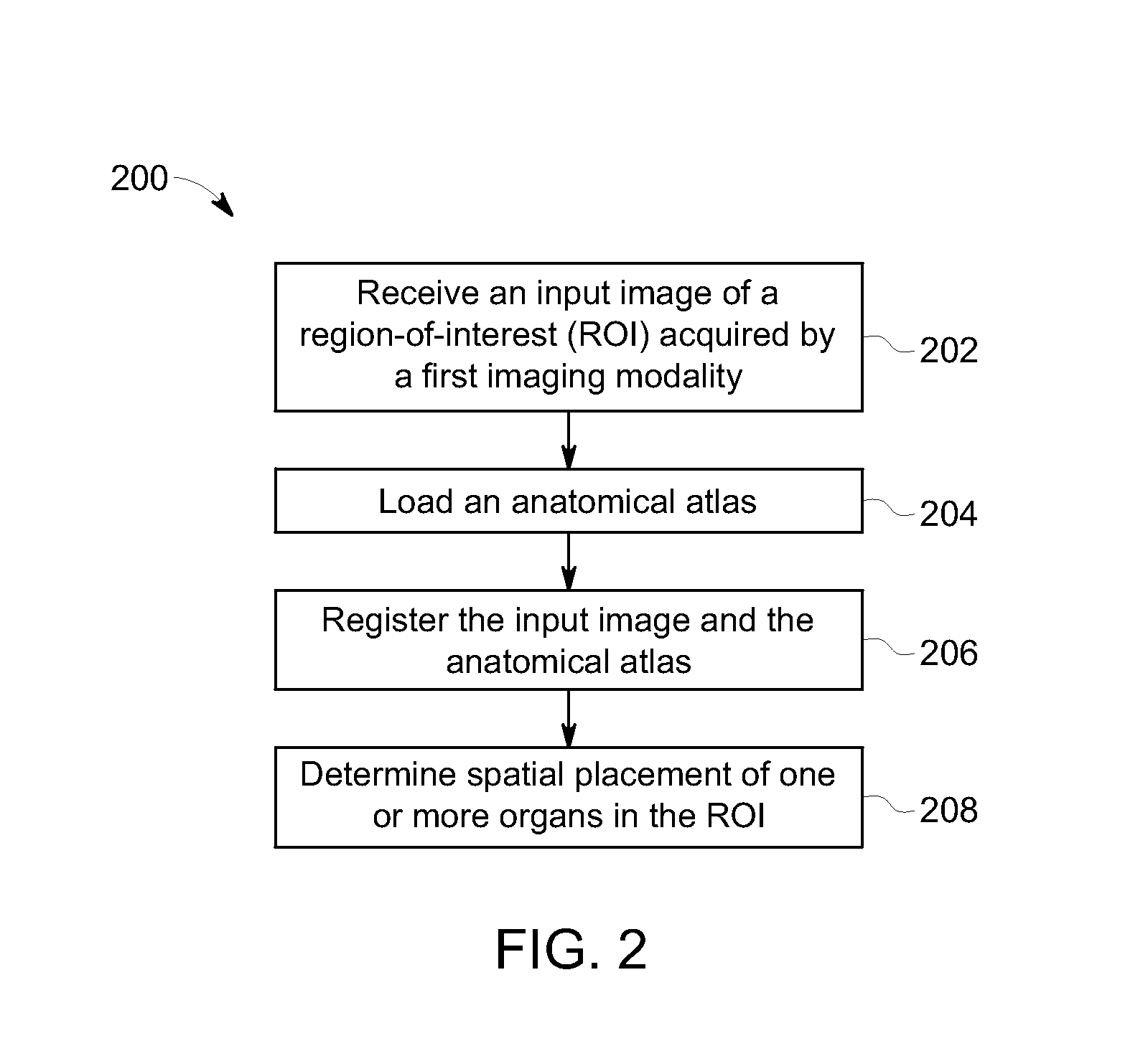
[0016]Embodiments described herein include methods, systems, and computer readable media that may facilitate at least one of processing or analyzing medical images of a region-of-interest (ROI) (also referred to as a volume-of-interest (VOI)). For example, embodiments may include methods, systems, and computer readable media that generate a multi-modality anatomical atlas. Embodiments may also include methods, systems, and computer readable media that determine a spatial placement of one or more organs in a region-of-interest (ROI).

[0017]The medical images may include image data or datasets that represent a visualization of the ROI. The image data may include pixels (or voxels) having signal intensity values or other values / qualities / characteristics that may be processed to form the visualization. Various imaging modalities may be used to acquire the medical images. Non-limiting examples of such modalities include ultrasound, magnetic resonance imaging (MRI), computed tomography (CT...


PUM


Abstract

A method that includes receiving an input image of a region of interest (ROI) of an individual. The input image is a medical image acquired by a first imaging modality. The method also includes generating a first feature image based on the input image. The first feature image includes a designated anatomical feature of the ROI. The method also includes obtaining an anatomical atlas. The atlas has a reference image of the ROI of at least one other individual and an organ model. The reference image is a medical image that is acquired by a second imaging modality that is different from the first imaging modality. The method also includes determining a transformation function by registering the first feature image with a second feature image that is based on the reference image and includes the designated anatomical feature.

Description

BACKGROUND[0001]The subject matter disclosed herein relates generally to medical imaging systems, and more particularly, to methods and systems that facilitate registering medical images for analysis.[0002]Various image-processing workflows exist in which medical images may undergo registration and segmentation operations. During a registration operation, medical images from different time periods and / or medical images acquired through different modalities are mapped together. During a segmentation operation, anatomical structures or features are extracted from the medical images. For example, a medical image may be analyzed to identify pixels (or voxels) of the medical images as corresponding to a designated organ (e.g., liver) or a designated tissue (e.g., soft tissue). The medical images may be acquired by different imaging modalities (e.g., ultrasound; magnetic resonance imaging (MRI); computed tomography (CT); positron emission tomography (PET); single photon emission computed ...

Claims


Application Information

Patent Timeline
Patent Type & Authority Patents(United States)
IPC IPC(8): G06K9/00G06T7/00
CPCG06T7/0028G06T2207/10072G06T2207/30004G06T7/33G06T7/70G06T2207/10081G06T2207/20221G06T2207/10108G06T2207/10076G06T2207/10084G06T2207/20048
Inventor KRISTON, ANDRASKOVACS, FERENCBLASKOVICS, TAMAS
Owner GENERAL ELECTRIC CO
Why Eureka
Try Eureka