Method for detecting nucleosome adducts

a nucleosome and nucleosome technology, applied in the field of nucleosome adduct detection, can solve the problems of tissue sample collection invasive involving surgery or biopsy, nucleosome and dna measurement in serum or plasma not agreeing well, and testing not agreeing with each other

Active Publication Date: 2015-11-17
SINGAPORE VOLITION PTE LTD
View PDF9 Cites 10 Cited by
  • Summary
  • Abstract
  • Description
  • Claims
  • Application Information

AI Technical Summary

Benefits of technology

[0193]In one embodiment, the method of the invention is repeated on multiple occasions. This embodiment provides the advantage of allowing the detection results to be monitored over a time period. Such an arrangement will provide the benefit of monitoring or assessing the efficacy of treatment of a disease state. Such monitoring methods of the invention can be used to monitor onset, progression, stabilisation, amelioration, relapse and / or remission.

Problems solved by technology

However, we have found that the results of these assays do not agree with each other.
Furthermore, although most circulating DNA in serum or plasma is reported to exist as mono-nucleosomes and oligo-nucleosomes (Holdenrieder et al, 2001), measured levels of nucleosomes and DNA in serum or plasma do not agree well.
One disadvantage of immunohistochemical methods for clinical use is that tissue sample collection is invasive involving surgery or biopsy.
These methods are well known in the art but are complex, laborious and expensive.

Method used


Image

  • Method for detecting nucleosome adducts
  • Method for detecting nucleosome adducts
  • Method for detecting nucleosome adducts

Examples


example 1

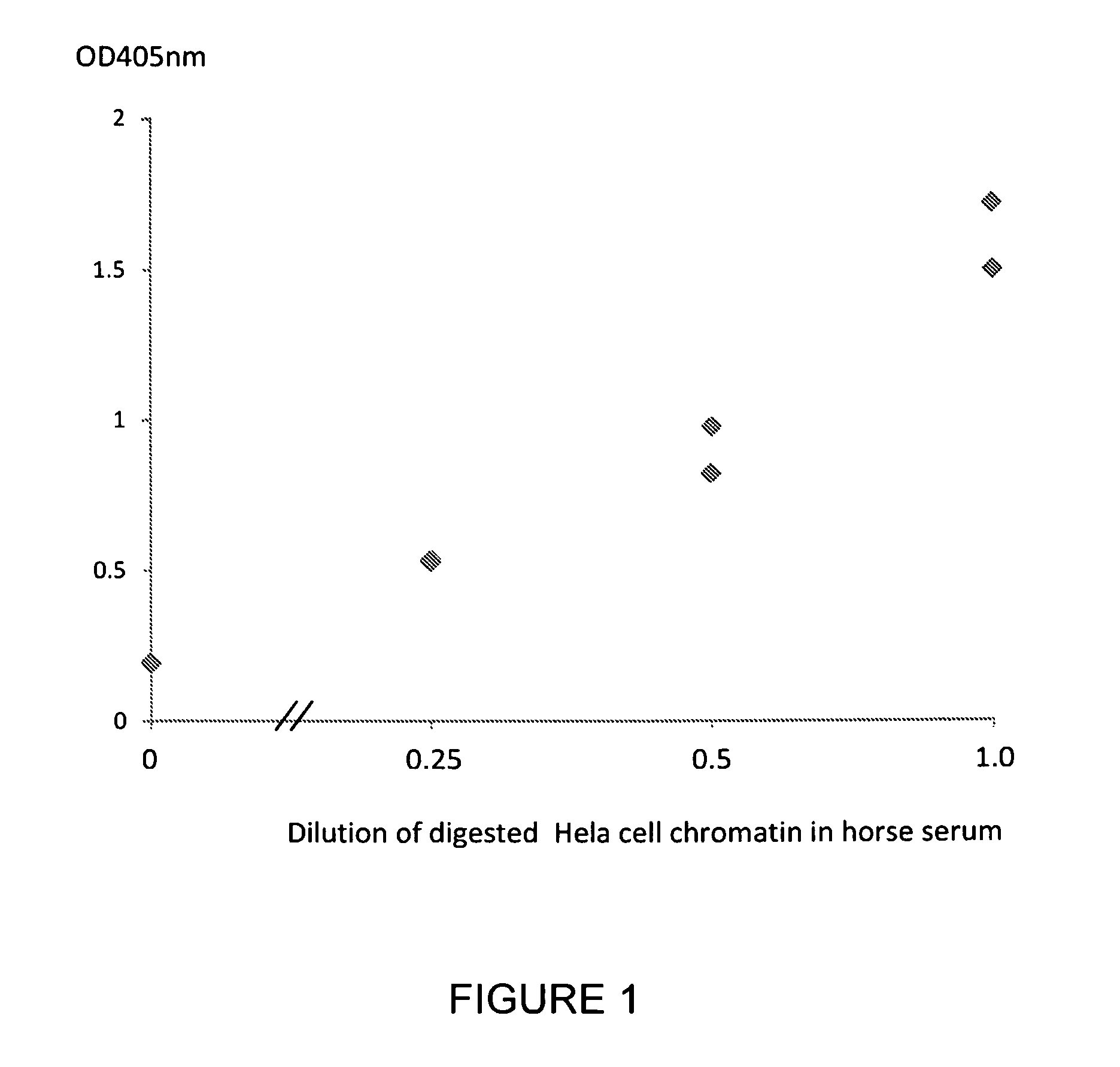
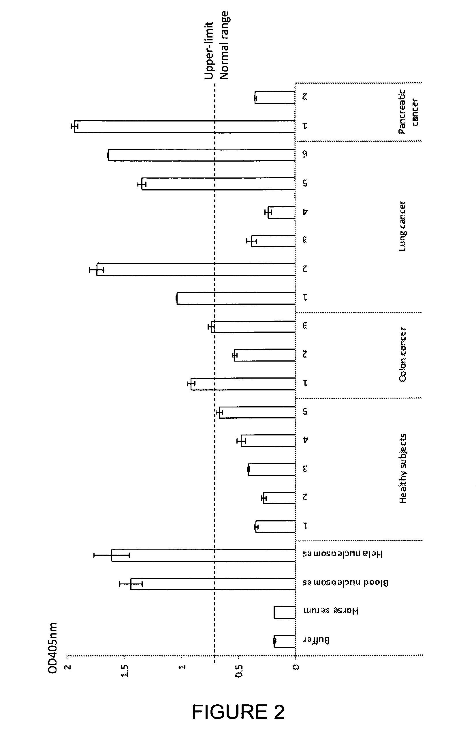
[0224]Serum samples were taken from 5 healthy subjects, 3 subjects with colon cancer, 6 subjects with lung cancer and 2 subjects with pancreatic cancer. A commercially available nucleosome preparation produced by digestion of chromatin extracted from Hela cells, in which the DNA and proteins in the nucleosome are cross-linked for stability, was serially diluted in horse serum. A nucleosome preparation in human blood was prepared according to the method of Holdenrieder (*Holdenrieder et al; 2001). These samples and preparations were assayed in duplicate for nucleosome-EZH2 adduct by the method of the invention. Neat commercially available horse serum produced for use in tissue culture was also assayed as a negative control sample containing no nucleosomes or nucleosome adducts.

[0225]The ELISA method used a solid phase anti-histone capture antibody that binds intact nucleosomes and a biotinylated monoclonal anti-EZH2 detection antibody as follows: A solution of anti-histone antibody i...

example 2

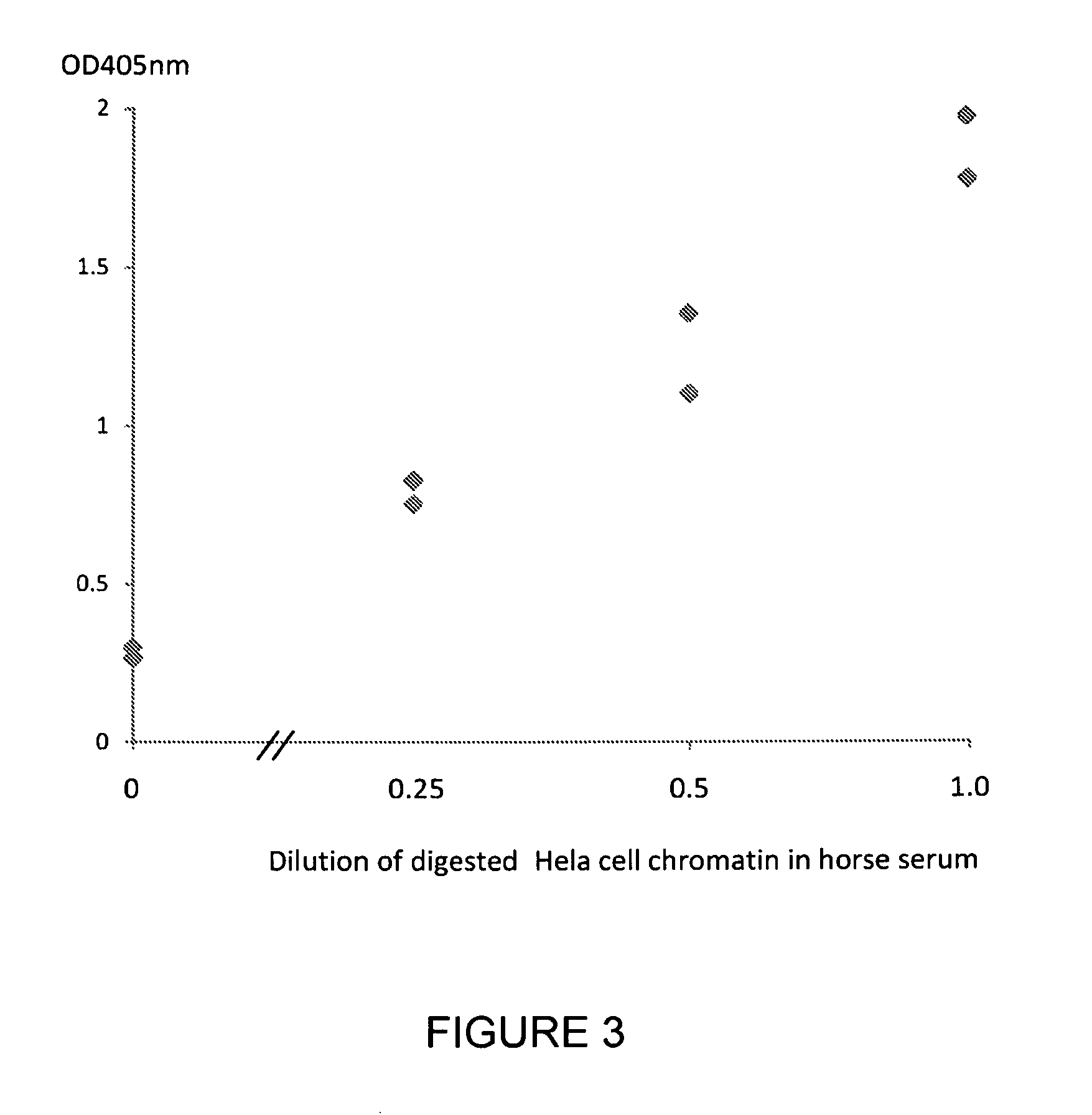
[0226]Serum samples were taken from 5 healthy subjects, 3 subjects with colon cancer, 6 subjects with lung cancer and 2 subjects with pancreatic cancer. A commercially available nucleosome preparation produced by digestion of chromatin extracted from Hela cells, was serially diluted in horse serum. A nucleosome preparation in human blood was prepared according to the method of Holdenrieder (*Holdenrieder of al; 2001). These samples and preparations were assayed in duplicate for nucleosome-HMGB1 adduct by the method of the invention. Neat horse serum was also assayed as a negative control sample containing no nucleosomes or nucleosome adducts.

[0227]The ELISA method used a solid phase anti-histone capture antibody that binds intact nucleosomes and a biotinylated monoclonal anti-HMGB1 detection antibody as follows: A solution of anti-histone antibody in 0.1M phosphate buffer pH 7.4 was added to microtitre wells (100 μL / well) and incubated overnight at 4° C. to coat the wells with captu...

example 3

[0233]A nucleosome-PR ELISA assay was carried out using the method of Example 1 above except that the biotinylated antibody used was directed to bind the progesterone receptor (PR). The results are shown in FIG. 6.


PUM

PropertyMeasurementUnit
Optical Densityaaaaaaaaaa
pHaaaaaaaaaa

Abstract

The invention relates to a method for detecting and measuring the presence of nucleosome-protein adducts and the use of such measurements for the detection and diagnosis of disease. The invention also relates to a method of identifying nucleosome adduct biomarkers for the detection and diagnosis of disease and to biomarkers identified by said method.

Description

FIELD OF THE INVENTION[0001]The invention relates to a method for detecting and measuring the presence of nucleosome-protein adducts and the use of such measurements for the detection and diagnosis of disease. The invention also relates to a method of identifying nucleosome adduct biomarkers for the detection and diagnosis of disease and to biomarkers identified by said method.BACKGROUND OF THE INVENTION[0002]The human body comprises several hundred cell types. All of these cell types contain the same genome but widely different phenotypes and different functions in the body. This phenotypic diversity is due to the differential expression of the genome in different cell types. The control of differential gene expression is not entirely understood but the basic mechanisms include gene regulation by a number of interconnected epigenetic signals associated with the gene, including control of the chromatin packing as euchromatin or heterochromatin, control of nucleosome positioning and ...

Claims


Application Information

Patent Timeline
Patent Type & Authority Patents(United States)
IPC IPC(8): G01N33/53G01N33/68G01N33/574C12Q1/68
CPCC12Q1/6804G01N33/57488G01N33/57496G01N33/6875G01N2800/52G01N2800/56G01N2800/7095G01N33/564G01N33/574G01N33/68G01N2333/47
Inventor MICALLEF, JACOB VINCENTECCLESTON, MARK EDWARDHERZOG, MARIELLE
Owner SINGAPORE VOLITION PTE LTD
Why Eureka
Try Eureka