
System and method for partial scan artifact reduction in myocardial CT perfusion

a computed tomography and reconstruction artifact technology, applied in the field of medical imaging, can solve the problems of difficult cardiac ct, inability to guarantee that the same angular data range (or view angle) is covered by each consecutive partial scan, and variations in the number of artifactual ct over time, and achieve the effect of introducing additional doses

Active Publication Date: 2016-01-12
MAYO FOUND FOR MEDICAL EDUCATION & RES
View PDF28 Cites 3 Cited by
  • Summary
  • Abstract
  • Description
  • Claims
  • Application Information

AI Technical Summary

Benefits of technology

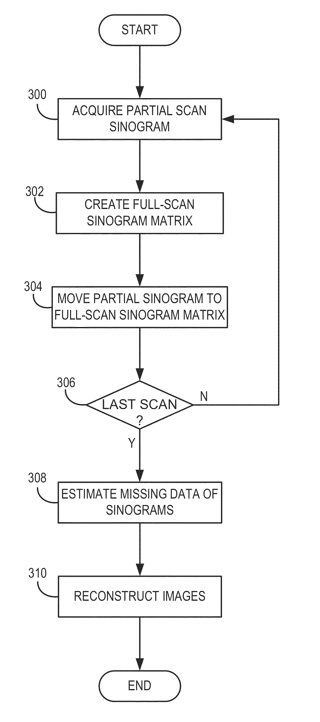
[0010]The present invention overcomes the aforementioned drawback by providing a system and method for processing and reconstructing partial-scan data acquired by a CT system without the need to use specialized hardware, introduce additional dose to the patient, or endure burdensome computations. Rather, the present disclosure provides a system and method for transforming partial-scan sinogram data into full-scan sinogram data and reconstructing images form the full-scan sinogram data, without the drawbacks of traditional methods for overcoming PSAs described above.
[0013]In accordance with yet another aspect of the disclosure, a method for reducing partial scan reconstruction artifacts in computed tomography images acquired during subject motion using partial scan trajectories is provided. The method includes obtaining at least one set of medical imaging projection data from a subject undergoing motion using partial computed tomography scans having an associated angular range and storing the at least one set of medical imaging projection data in a matrix arranged according to view angle. The method also includes estimating medical imaging projection data in view angles outside the angular range covered by each partial computed tomography scan using at least one of a weighting and an interpolation based on the medical imaging projection data from neighboring partial computed tomography scans.

Problems solved by technology

However, from a technical perspective, cardiac CT is challenging due to demands in temporal resolution.
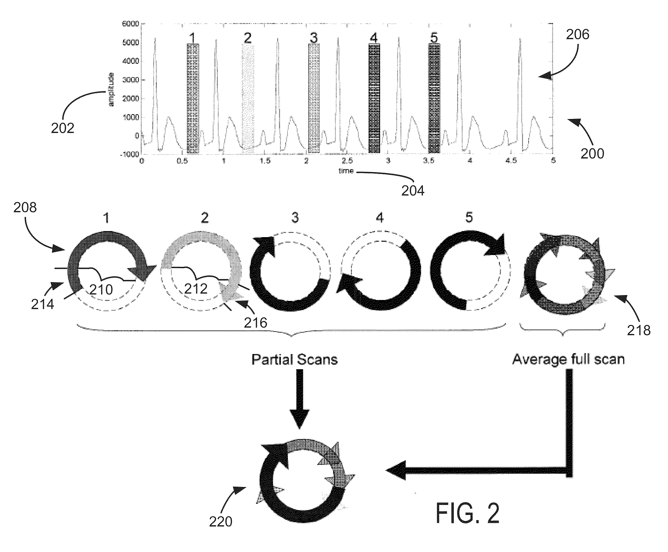
Since myocardial CTP protocols are typically ECG-gated, according to a desired interval within the cardiac cycle (for example, diastolic phase), it is not possible to guarantee that the same angular data range (or view angles) is covered by each consecutive partial scan.
As a consequence, artifactual CT number variations over time, called partial scan reconstruction artifacts (PSR artifacts or PSAs), arise due to minor deviations in the projection data due to physical effects, for example, such as scattering and beam hardening.
However, the obvious disadvantage of such an approach is its invasiveness, which precludes its use in humans.
A disadvantage of this approach is an increase in radiation dose (for example, 14%), and potential decreases in temporal resolution compared to a partial scan.
A disadvantage is that the method requires several image reconstruction steps (3 instead of 1); hence, computational requirements are increased at least threefold.
Another disadvantage is that, in practice, the superposition operations are potentially error prone because of mismatches generated by motion artifacts due to the rapid heart motion, that is, when applying this technique in vivo.

Method used


Image

  • System and method for partial scan artifact reduction in myocardial CT perfusion
  • System and method for partial scan artifact reduction in myocardial CT perfusion
  • System and method for partial scan artifact reduction in myocardial CT perfusion

Examples


Embodiment Construction

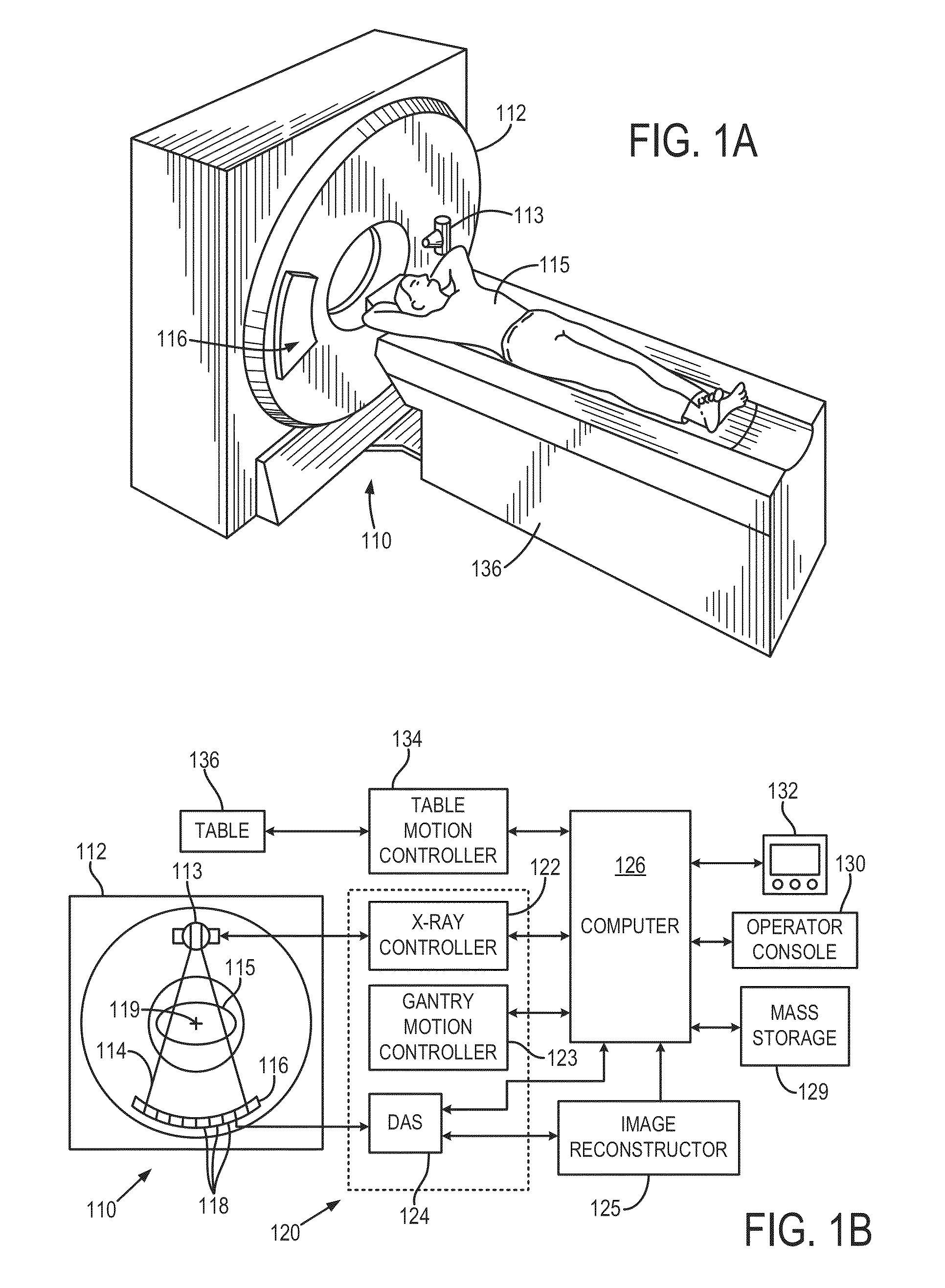
[0020]With initial reference to FIGS. 1A and 1B, an x-ray computed tomography (“CT”) imaging system 110 includes a gantry 112 representative of a “third generation” CT scanner. Gantry 112 has an x-ray source 113 that projects a fan-beam, or cone-beam, of x-rays 114 toward a detector array 116 on the opposite side of the gantry. The detector array 116 is formed by a number of detector elements 118 which together sense the projected x-rays that pass through a medical patient 115. Each detector element 118 produces an electrical signal that represents the intensity of an impinging x-ray beam and hence the attenuation of the beam as it passes through the patient. During a scan to acquire x-ray projection data, the gantry 112 and the components mounted thereon rotate about a center of rotation 119 located within the patient 115.

[0021]The rotation of the gantry and the operation of the x-ray source 113 are governed by a control mechanism 120 of the CT system. The control mechanism 120 inc...


PUM


Abstract

A system and method is provided for reducing partial scan reconstruction artifacts in sinogram data acquired as a series of sets of partial-scan projection views, each set of projection views extending over an angular range of less than 360 degrees. A full-scan sinogram matrix for each of the sets of projection views in the series is created and each set of partial-scan projection views is stored in a respective full-scan sinogram matrix to create an array of full-scan matrices having respective empty spaces not filled by partial-scan projection view data stored therein. The empty spaces are filled in each of the full-scan sinogram matrices using the partial-scan projection view data stored therein and an image of the subject is reconstructed from the full-scan sinogram matrices having the empty spaces filled using the partial-scan projection view data stored therein.

Description

CROSS-REFERENCE TO RELATED APPLICATIONS[0001]This application is based on, claims the benefit of, and incorporates herein by reference U.S. Provisional Patent Application Ser. No. 61 / 613,475, entitled, filed Mar. 20, 2012, and entitled “SYSTEM AND METHOD FOR PARTIAL SCAN ARTIFACT REDUCTION IN MYOCARDIAL CT PERFUSION” and also U.S. Provisional Patent Application Ser. No. 61 / 625,415, entitled, filed Apr. 17, 2012, and entitled “SYSTEM AND METHOD FOR PARTIAL SCAN ARTIFACT REDUCTION IN MYOCARDIAL CT PERFUSION”STATEMENT REGARDING FEDERALLY SPONSORED RESEARCH[0002]N / ABACKGROUND OF THE INVENTION[0003]The present invention relates generally to systems and methods for medical imaging and, particularly, to systems and methods for reducing partial scan reconstruction artifacts in computed tomography perfusion (CTP).[0004]In a computed tomography (“CT”) system, an x-ray source projects a fan-shaped or cone-shaped beam of x-rays that is collimated to lie within an x-y plane of a Cartesian coordi...

Claims


Application Information

Patent Timeline
Patent Type & Authority Patents(United States)
IPC IPC(8): G06K9/00A61B6/00G06T11/00A61B6/03
CPCG06T11/003A61B6/032A61B6/481A61B6/486A61B6/487A61B6/503A61B6/507A61B6/5264A61B6/541G06T11/006G06T2211/412G06T2211/432G06T2211/436
Inventor RAMIREZ GIRALDO, JUAN C.MCCOLLOUGH, CYNTHIA H.
Owner MAYO FOUND FOR MEDICAL EDUCATION & RES
Learn More