

Methods for detecting antibodies
a detection method and antibody technology, applied in the field of detection of antibodies, can solve the problems of large-scale sample analysis and easy automation of assays
- Summary
- Abstract
- Description
- Claims
- Application Information
AI Technical Summary
Benefits of technology
Problems solved by technology
Method used
Image

Examples
example 1
[0086]While the following assay reagent protocol is described using specifically identified reagents, such as specific fluorescent labels, specific phage-displayed libraries and generation of specifically identified peptides, the methods described herein may be utilized to generate peptides including mimetopes that interact with any antibody.
[0087]Antibodies were prepared as follows. Alemtuzumab (Genzyme, Cambridge, Mass.) and rituximab (Genentech, San Francisco, Calif.) were obtained from the UCSD Cancer Center pharmacy. The antibodies were fluorescently labeled using the Zenon® R-Phycoerythrin Human IgG labeling kit, Zenon® Alexa Fluor® 488 Human IgG labeling and Zenon® Alexa Fluor® 488 protein labeling kits (Invitrogen, Carlsbad, Calif.). For kinetic studies, Fab fragments of each mAb were prepared by papain digestion using a Fab preparation kit (Thermo Fisher Scientific, Rockford, Ill.) as per manufacturers instructions.
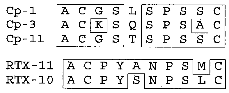
[0088]Peptide mimetopes were gener...
example 2
Binding Kinetics Analysis Protocol
[0092]Kinetics of antibody-peptide binding were studied using SkiPro™ Biomolecular Interaction technology platform on a SkiPro™ interferometer equipped with a 2-channel flow cell and an autosampler (Silicon Kinetics Inc. San Diego, Calif.). All reagents were purchased from Sigma-Aldrich (St. Louis, Mo.). All peptides were incubated with oxidizing agent Sodium Tetrathionate at 10 mM in PBS for 1 hour immediately before binding experiments. Biotinylated peptides were diluted to 5 μM in PBS / 0.05% BSA and immobilized on Streptavidin-coated SkiSensor™ Biochips for 10 minutes at 4 μl / min flow rate. That resulted in an Optical Path Difference shift (ΔOPD) of 5-6 nm. Concentration series of antibodies and antibody Fab fragments were prepared as twofold dilutions in PBS / 0.05% BSA. Binding was carried out for 10 minutes followed by 20 minutes dissociation. For all sensorgrams reference channel data was subtracted from the sample channel. The resulting multi-c...
example 3
Detection of Antibodies in Chronic Lymphoid Leukemia Cells
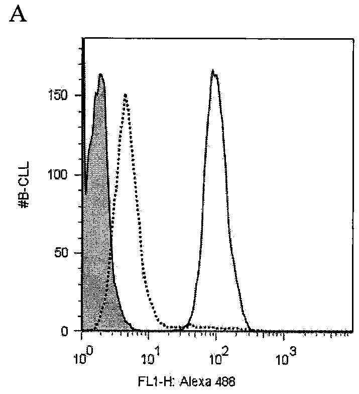
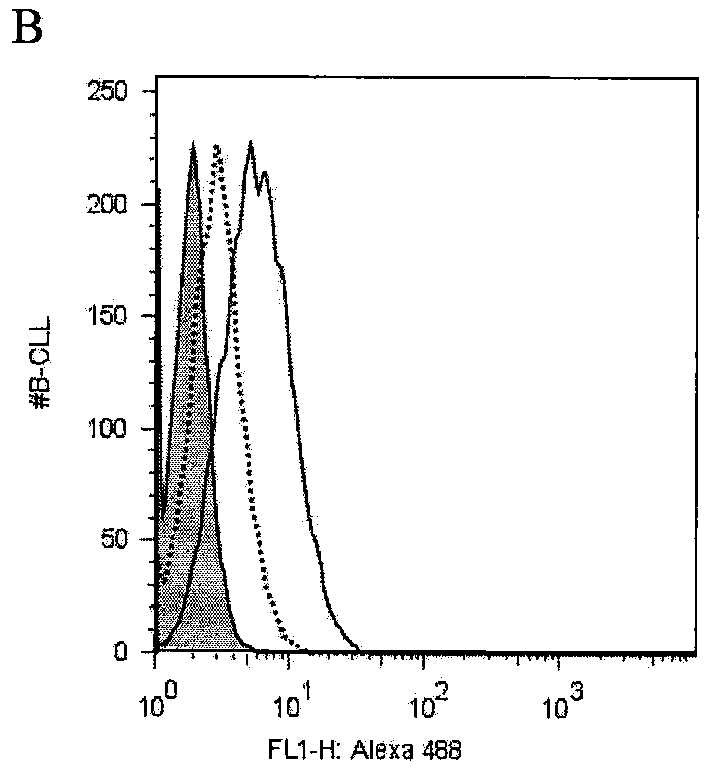
[0093]This example illustrates detection of antibodies (alemtuzumab and rituximab) in chronic lymphoid leukemia (CLL) cells and serum samples. The cells were used in blocking experiments to determine the ability of peptide mimetopes specific for the antibodies (alemtuzumab and rituximab) to inhibit the binding of the antibodies to the surface of the primary CLL cells.
[0094]After informed consent was obtained per the Declaration of Helsinki, blood samples were collected from patients at the University of California, San Diego (UCSD) Medical Center who satisfied diagnosis and immunophenotypic criteria for common B-cell CLL.
[0095]CLL cells (105 / per well) were seeded into 96-well plates in 100 μl of X-vivo™ medium (BioWhittaker, Walkersville, Md.), and incubated with Zenon® labeled alemtuzumab or rituximab prepared as described in Example I (0.5 μg / well) for 1 h on ice. Subsequently, cells were washed two times with 100 μl FACS-w...
PUM
| Property | Measurement | Unit |
|---|---|---|
| concentrations | aaaaa | aaaaa |
| concentrations | aaaaa | aaaaa |
| particle size | aaaaa | aaaaa |
Abstract
Description
Claims
Application Information