
Method and apparatus for real-time and robust strain imaging

a real-time and robust technology, applied in image enhancement, instruments, ultrasonic/sonic/infrasonic image/data processing, etc., can solve the problems of many false positive alarms, significant unnecessary costs and psychological burdens, and unnecessarily selecting benign lesions for biopsy, etc., to achieve the effect of improving robustness

Active Publication Date: 2017-06-27
SHARAFAT AHMAD R +1
View PDF8 Cites 0 Cited by
  • Summary
  • Abstract
  • Description
  • Claims
  • Application Information

AI Technical Summary

Benefits of technology

The present invention provides a way to see strain in real-time during elastography, while being more robust against variations in compression caused by operator's hand movements.

Problems solved by technology

However, false positive alarms are common in screening, leading to significant unnecessary costs and psychological burdens in such cases.
However, similarities between physical and mammographic symptoms of benign and malignant lesions result in many false positive alarms.
In such cases, benign lesions are unnecessarily chosen for biopsy.
But due to practical limitations for medical imaging, only ultrasound and magnetic resonance imaging (MRI) systems have been utilized for this purpose so far.
The cost saving in freehand elastography comes at the expense of some limitations.
However, the tendency of objects for out-of-plane motion makes it difficult to perform such compression, especially for deeper organs that are less influenced by the applied stress.
These limitations can greatly degrade the quality of strain maps.
However, this approach is too time-consuming for busy clinical practice.
However, due to the fact that calculations are minimized in real-time processing, real-time algorithms may suffer from lack of quality and robustness as compared with offline strain imaging methods.
More specifically, they are more vulnerable to “signal decorrelation”, which is defined as the lack of correlation between pre- and post-deformation signals, caused by increasing the applied stress.
However, the decorrelation issue at the first step of the algorithm could affect the entire displacement estimation process in this approach.
Although the regularization enhances the robustness of the algorithm against signal decorrelation, this issue can still affect the accuracy of the results through the seed line, where inaccurate estimation would spread to the whole image.

Method used


Image

  • Method and apparatus for real-time and robust strain imaging
  • Method and apparatus for real-time and robust strain imaging
  • Method and apparatus for real-time and robust strain imaging

Examples


Embodiment Construction

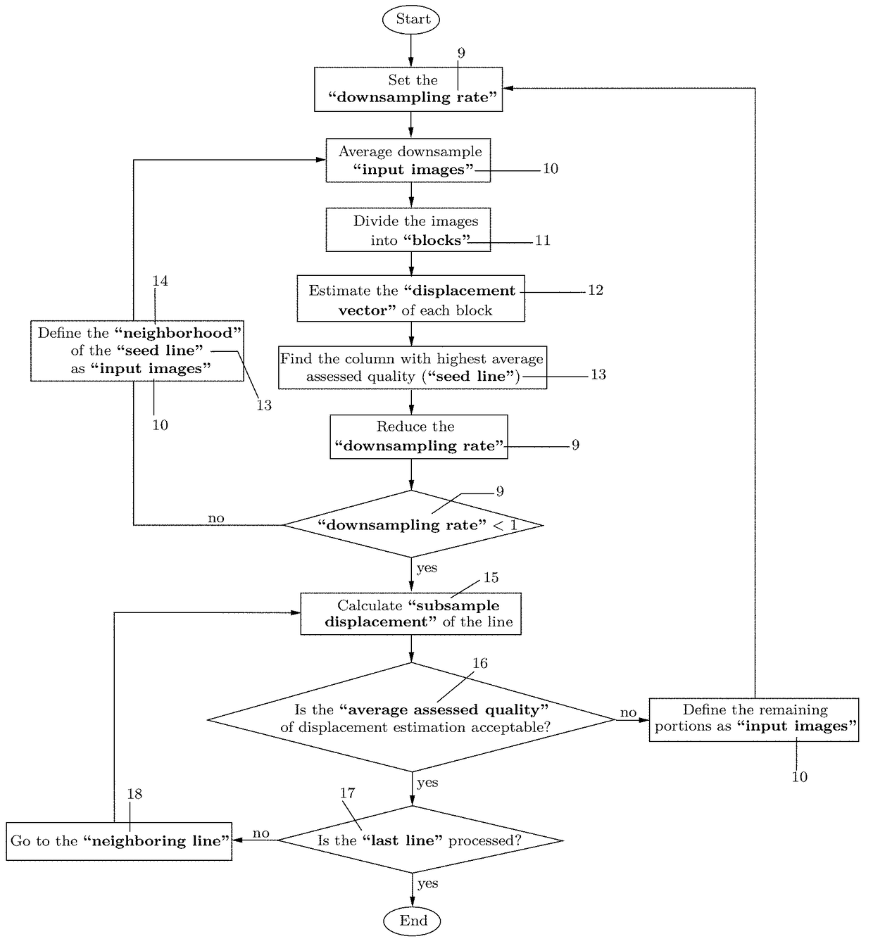
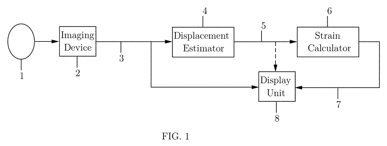
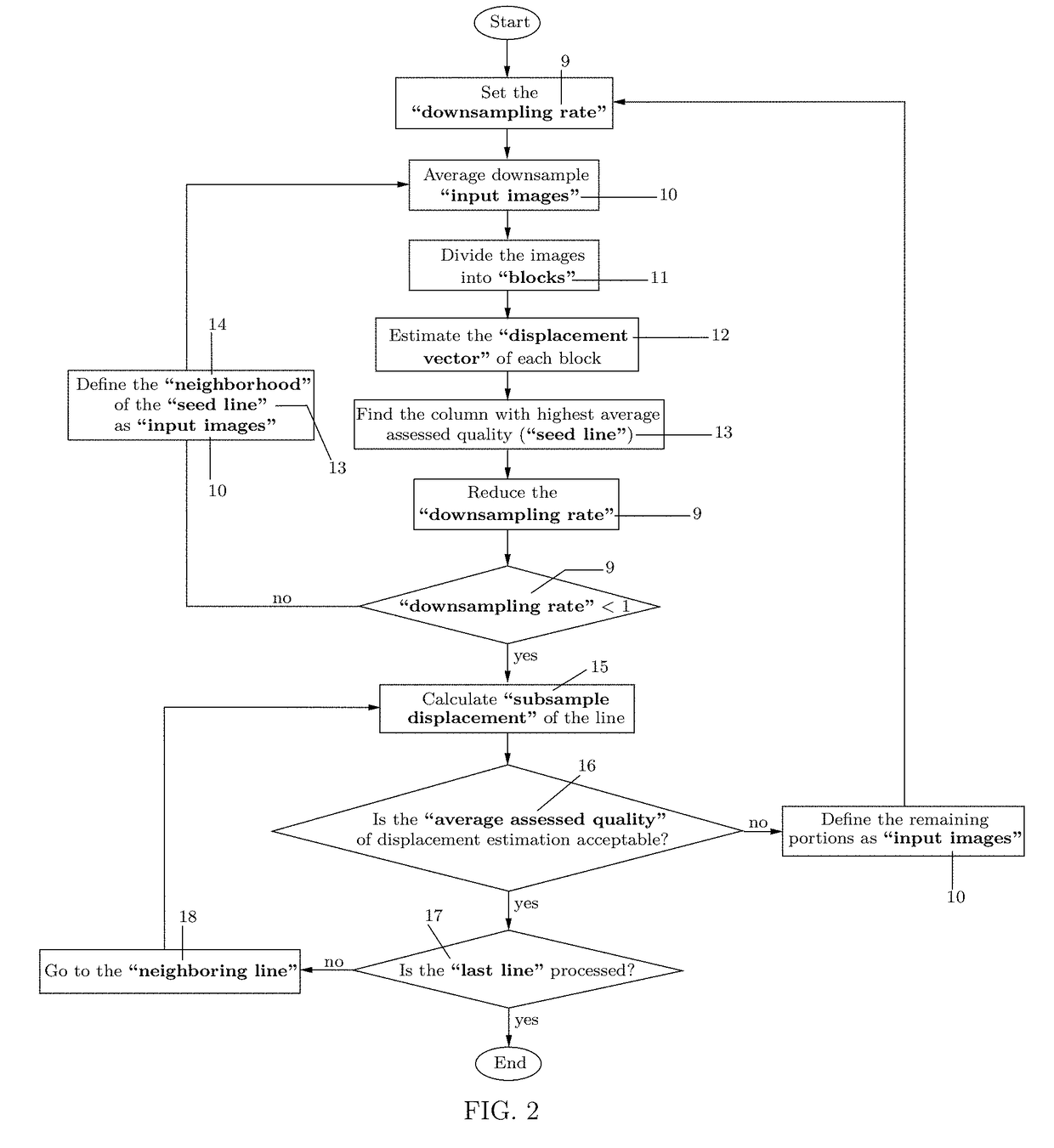
[0044]FIG. 1 shows a high-level block diagram of the strain map formation process of an object [1]. An imaging device [2] provides pre- and post-deformation images [3] of pre- and post-deformation states of the object [1]. The deformation may be performed by an external compression fixture, such as a mechanical arm; or a part of the imaging device [2] can be used for compressing the object [1], such as the transducer of ultrasound imaging system. In some applications, natural compression sources such as arterial pulsation, cardiac motion, or patient breathing may be utilized for object compression.

[0045]Theoretically, the imaging device [2] can be any type of imaging modalities. Currently, ultrasound imaging is most commonly practiced for elasticity imaging, and some preferred embodiments of the present invention are described with reference to ultrasonic data processing. However, other imaging systems such as, potentially, magnetic resonance imaging and computerized tomography may ...


PUM


Abstract

A robust algorithm is disclosed for real-time strain imaging. The method can be implemented on conventional medical imaging modalities such as ultrasound, MRI, etc. The pre- and post-deformation images, acquired by an imaging system, are used by the algorithm to produce the strain map. Deformation can be performed by any available means, such as an existing part of the imaging device (as in freehand elastography) or a separate compression fixture. Due to the computational efficiency of the algorithm, any conventional processing platform, including personal computers, tablet PCs, and smart phones can be employed for strain imaging, to construct an ultra-portable strain imaging system.

Description

TECHNICAL FIELD[0001]Elasticity imaging, medical imaging.BACKGROUND OF THE INVENTION[0002]Cancer is a leading cause of death in the world. According to the World Health Organization (WHO), about 8.2 million people died because of cancer in 2012, 70% of whom were living in developing countries. The number of cancer cases is expected to rise from 14 million in 2012 to 22 million in the next two decades.[0003]Screening is widely accepted as an effective strategy for early detection of cancer, which can increase the rate of successful treatment. However, false positive alarms are common in screening, leading to significant unnecessary costs and psychological burdens in such cases. One major instance is screening the breast cancer, which is the most fatal cancer among women, causing more than 500,000 deaths worldwide in 2012.[0004]Breast cancer screening is usually performed via mammography and clinical breast examination. However, similarities between physical and mammographic symptoms ...

Claims


Application Information

Patent Timeline
Patent Type & Authority Patents(United States)
IPC IPC(8): A61B8/08A61B8/14G06T7/00G06K9/46A61B8/00A61B5/00A61B5/055
CPCA61B8/485A61B8/14A61B8/403A61B8/463A61B8/5223G06K9/4604G06T7/0012G06T7/231A61B5/055A61B5/7282A61B5/7425G06T2207/10088G06T2207/10132G06T2207/30096G06T7/0016G06T2207/10016G06T2207/20016G06T2207/20021G06T2207/30068G06T7/223A61B8/085A61B8/4427A61B8/5207A61B8/587
Inventor SHARAFAT, AHMAD R.REZAJOO, SAEED
Owner SHARAFAT AHMAD R
Learn More