Method and apparatus to determine a magnetic resonance relaxation time in the heart muscle in a magnetic resonance examination

a magnetic resonance examination and relaxation time technology, applied in the field of methods to determine the relaxation time of the heart muscle, can solve the problem that the prior art-based inline segmentation algorithm cannot automatically d

Active Publication Date: 2018-04-24
SIEMENS HEALTHCARE GMBH
View PDF18 Cites 0 Cited by
  • Summary
  • Abstract
  • Description
  • Claims
  • Application Information

AI Technical Summary

Benefits of technology

[0014]According to a further embodiment of the method, the width of the annular slice image region is reduced in the radial direction in that the inner circumference of the slice region is enlarged by a predetermined value and the outer circumference of the slice region is reduced by a predetermined value. The predetermined value can be a value in the range from 1-10 mm, for example, or can be a predetermined percentile of 5%-30% of the entire width of the annular region, for example. Partial volume effects in the border region of the ventricle can be avoided by this narrowing of the annular slice image region, so the reliability of the method is improved.

Problems solved by technology

However, the inline segmentation algorithm known according to the prior art cannot automatically detect the septal region, which would be suitable as a reference tissue region to determine the T2* time, for example.

Method used


Image

  • Method and apparatus to determine a magnetic resonance relaxation time in the heart muscle in a magnetic resonance examination
  • Method and apparatus to determine a magnetic resonance relaxation time in the heart muscle in a magnetic resonance examination
  • Method and apparatus to determine a magnetic resonance relaxation time in the heart muscle in a magnetic resonance examination

Examples


Embodiment Construction

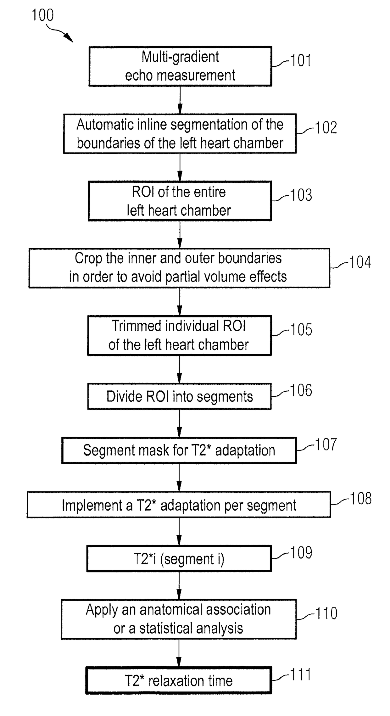
[0025]An MR system 700 with which an MR relaxation time in the heart muscle can be determined according to the invention in a magnetic resonance examination is shown in FIG. 7. The MR system 700 has a basic field magnet 701 that generates a polarization field B0. An examination person 703 arranged on a bed 702 is driven into the center of the basic field magnet 701 where the acquisition of the MR signals from an examination region is implemented via radiation of RF pulses and switching of gradients. How MR images—in particular images of a multi-gradient echo measurement—can be generated in a pulse sequence with a series of RF pulses and switching of gradients is known to those skilled in the art and need not be described in detail herein. The MR system 700 is connected with a central control unit 704 with which the MR system 700 is controlled. Among other things, the central control unit 704 has an RF control unit 705 which controls the switching of the RF pulses to deflect the magn...


PUM


Abstract

In a method and magnetic resonance (MR) system to determine an MR relaxation time (for example a T1, T2 or T2* relaxation time) in the heart muscle in a magnetic resonance examination, a determination an annular slice image region of the heart muscle of the left heart chamber in MR image data with the use of an automatic image segmentation. Multiple sub-regions within the slice image region are automatically determined. Each sub-region respectively includes multiple pixels of the annular slice image region of the heart muscle of the left heart chamber. An MR relaxation time is determined automatically for each of the multiple sub-regions and associated with the corresponding sub-region. A characteristic MR relaxation time in the heart muscle is determined by a statistical analysis of the multiple MR relaxation times that are associated with the multiple sub-regions.

Description

BACKGROUND OF THE INVENTION[0001]Field of the Invention[0002]The present invention concerns a method to determine a magnetic resonance (MR) relaxation time, in particular a T2* relaxation time, in the heart muscle in a magnetic resonance examination.[0003]Description of the Prior Art[0004]A determination of the MR relaxation times—for example a relaxation time T2*, a T1 relaxation time or a T2 relaxation time—is helpful in a number of magnetic resonance examinations.[0005]For example, a determination of the transversal relaxation time T2* is appropriate in magnetic resonance measurements (MR) for a diagnosis of thalassemia. Thalassemia is a genetically caused abnormality in hemoglobin production. Left untreated, patients can possibly suffer from anemia that is accompanied by an increased iron concentration in the heart muscle (myocardium). Evidence of the iron concentration in the myocardium is primarily relevant for therapy. A change in the iron concentration results in a shortenin...

Claims


Application Information

Patent Timeline
Patent Type & Authority Patents(United States)
IPC IPC(8): A61B5/05G01R33/50A61B5/055G01R33/24G01R33/56G01R33/561
CPCG01R33/50A61B5/055G01R33/24G01R33/5617G01R33/5608G01R33/5616G01R33/5602
Inventor GREISER, ANDREAS
Owner SIEMENS HEALTHCARE GMBH
Why Eureka
Try Eureka