Paralleled imaging method and system for common path type endoscopic OCT of hard tube model

An imaging system and tubular technology, applied in endoscopy, medical science, diagnosis, etc., can solve the problems of complex scanning mechanism and longer imaging time, and achieve the effect of fast imaging speed and simple operation

Inactive Publication Date: 2007-12-26
ZHEJIANG UNIV
View PDF0 Cites 15 Cited by
  • Summary
  • Abstract
  • Description
  • Claims
  • Application Information

AI Technical Summary

Problems solved by technology

Chen's group (Tuqiang Xie, et al., GRIN lensrod based probe for endoscopic spectral domain optical coherence tomography with fast dynamic focus tracking, Optics Express, 2006, 14(8): 3238-3246) proposed a GRIN lens rod based probe , gradient refractive index rod lens) hard-tube endoscopic probe, but placed on the sample arm of the non-common path interference structure, has the same problem as the above-mentioned endoscopic OCT system
In addition, due to the use of horizontal point scanning, the system requires a complex scanning mechanism, longer imaging time, and image reconstruction to obtain two-dimensional or three-dimensional images

Method used


Image

  • Paralleled imaging method and system for common path type endoscopic OCT of hard tube model
  • Paralleled imaging method and system for common path type endoscopic OCT of hard tube model

Examples


Embodiment Construction

[0031] Below in conjunction with accompanying drawing and embodiment the present invention will be further described:

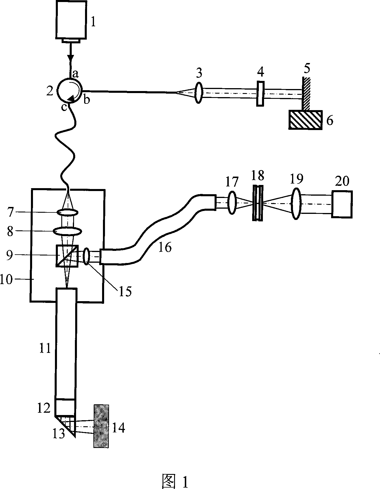
[0032] The hard-tube type common path endoscopic OCT parallel imaging system proposed by the present invention is shown in Figure 1. The light emitted by the broadband light source 1 is connected to the port a of the optical circulator 2, and then emitted to the first collimator by the port b of the optical circulator The lens 3 is divided into reflected light and transmitted light when collimated and incident on the broadband spectroscopic flat plate 4 in parallel, and the transmitted light is parallel incident on the mirror 5 fixed on the electronically controlled translation stage 6 . The mirror 5 adopts a wide-band high-reflectivity mirror surface, specifically, a metal-dielectric film broadband high-reflectance mirror, so as to ensure a high energy utilization rate of the system.

[0033]The light beam reflected by the mirror 5 and the broadband beam spl...


PUM


Abstract

The present invention discloses a hard-tube common-path type endoscopic OCT parallel imaging method and system. Said invention adopts Green rod lens with integer multiple cyclic length as endoscopic probe light-transferring device, uses column lens to implement linear focusing illumination of internal cavity wall, and utilizes face-matrix CCD detector to make detection of the spectral field signal. The font end face of the Green rod lens is placed on the rear focal plane of column lens, the integer multiple cyclic length can ensure that the light-beam form transferred to rear end face is identical to that incident upon front end face, so that the rear end face com be used as reference face, it and sample have conjugate imaging relationship, both they can be formed into a common-path sensitive interferometer. Said invention utilizes the combination of linear illumination parallel imaging technique and spectral field detection technique to obtain two-dimensional image of internal cavity wall.

Description

technical field [0001] The invention relates to medical endoscopic imaging technology and optical coherence tomography imaging technology, in particular to a hard-tube common-channel endoscopic OCT parallel imaging method and system using a Green rod lens as an endoscopic probe light transmission device. Background technique [0002] Medical endoscope is a non-invasive or minimally invasive medical instrument, with which doctors can directly observe the tissue lesions of internal organs of the human body and perform surgical treatment. Traditional medical endoscopes can only observe lesions on the surface of internal organs, and lesions often first occur in the faults of organs. The introduction of ultrasound technology enables the endoscope to observe the tissue morphology of organ slices while observing the mucosal surface of the lumen. However, its resolution is on the order of millimeters, which is far from being able to detect tissue abnormalities, such as the tissue st...

Claims


Application Information

Patent Timeline
Patent Type & Authority Applications(China)
IPC IPC(8): A61B1/055A61B1/05A61B1/002
Inventor 丁志华杨亚良吴兰
Owner ZHEJIANG UNIV
Why Eureka
Try Eureka