Rebuilding method of blood vessel three-dimensional model

A three-dimensional model and three-dimensional reconstruction technology, applied in the field of medical detection, can solve the problems of not considering the bending and twisting of the catheter, inaccurate results, accurate location and shape of lesions, etc.

Inactive Publication Date: 2008-10-15
NORTH CHINA ELECTRIC POWER UNIV (BAODING)
View PDF0 Cites 67 Cited by
  • Summary
  • Abstract
  • Description
  • Claims
  • Application Information

AI Technical Summary

Problems solved by technology

Since IVUS itself cannot provide spatial geometric information for each frame of image, this method does not take into account the bending and twisting of the catheter during image acquisition, and the results are also inaccurate
[0006] In short, there is currently no more accurate three-dimensional ...

Method used


Image

  • Rebuilding method of blood vessel three-dimensional model
  • Rebuilding method of blood vessel three-dimensional model
  • Rebuilding method of blood vessel three-dimensional model

Examples


Embodiment Construction

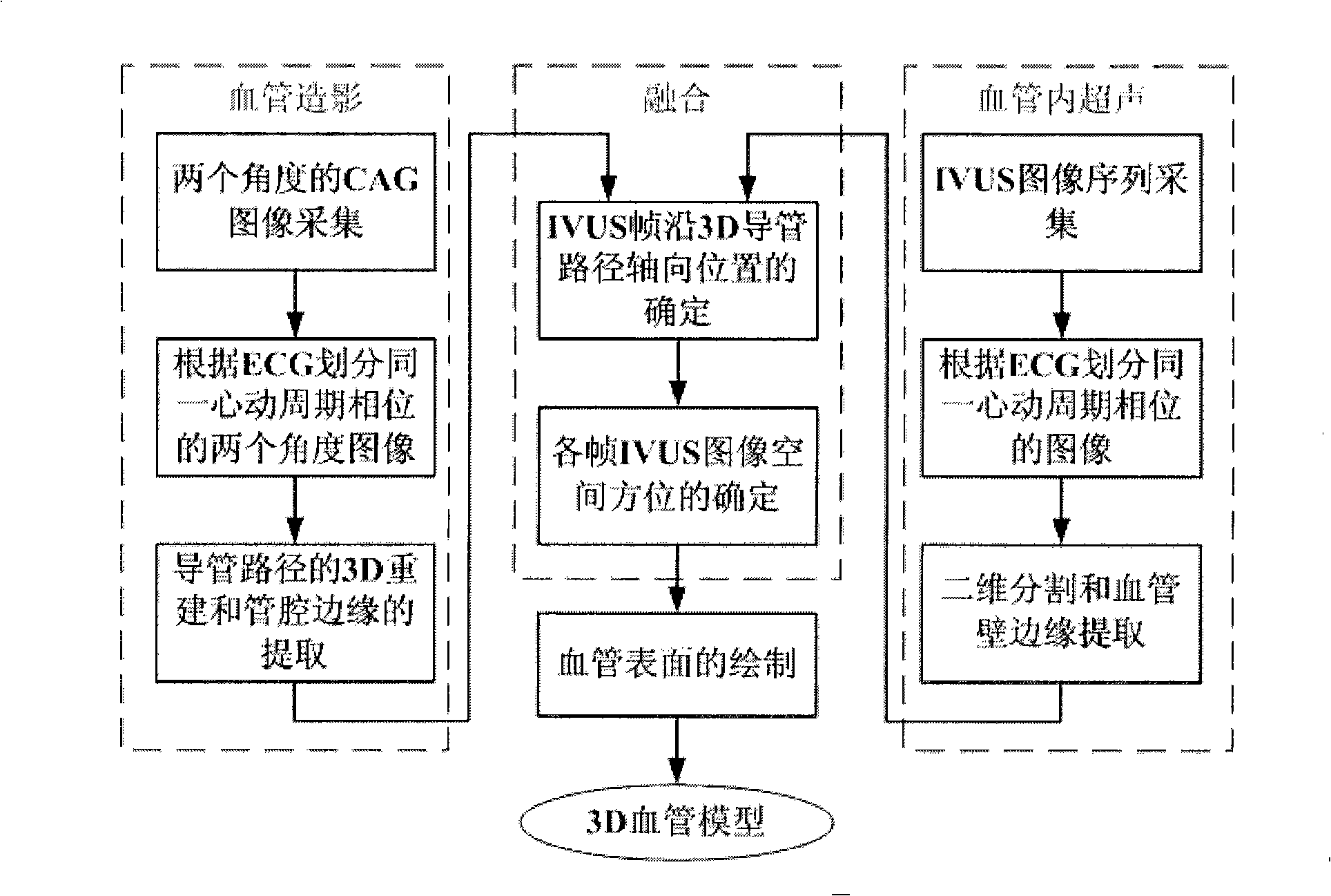
[0040] The steps of the present invention are described in detail below in conjunction with accompanying drawings:

[0041] (1) Image acquisition:

[0042] Acquisition equipment includes a C-arm single-sided X-ray angiography machine and an intravascular ultrasound imager.

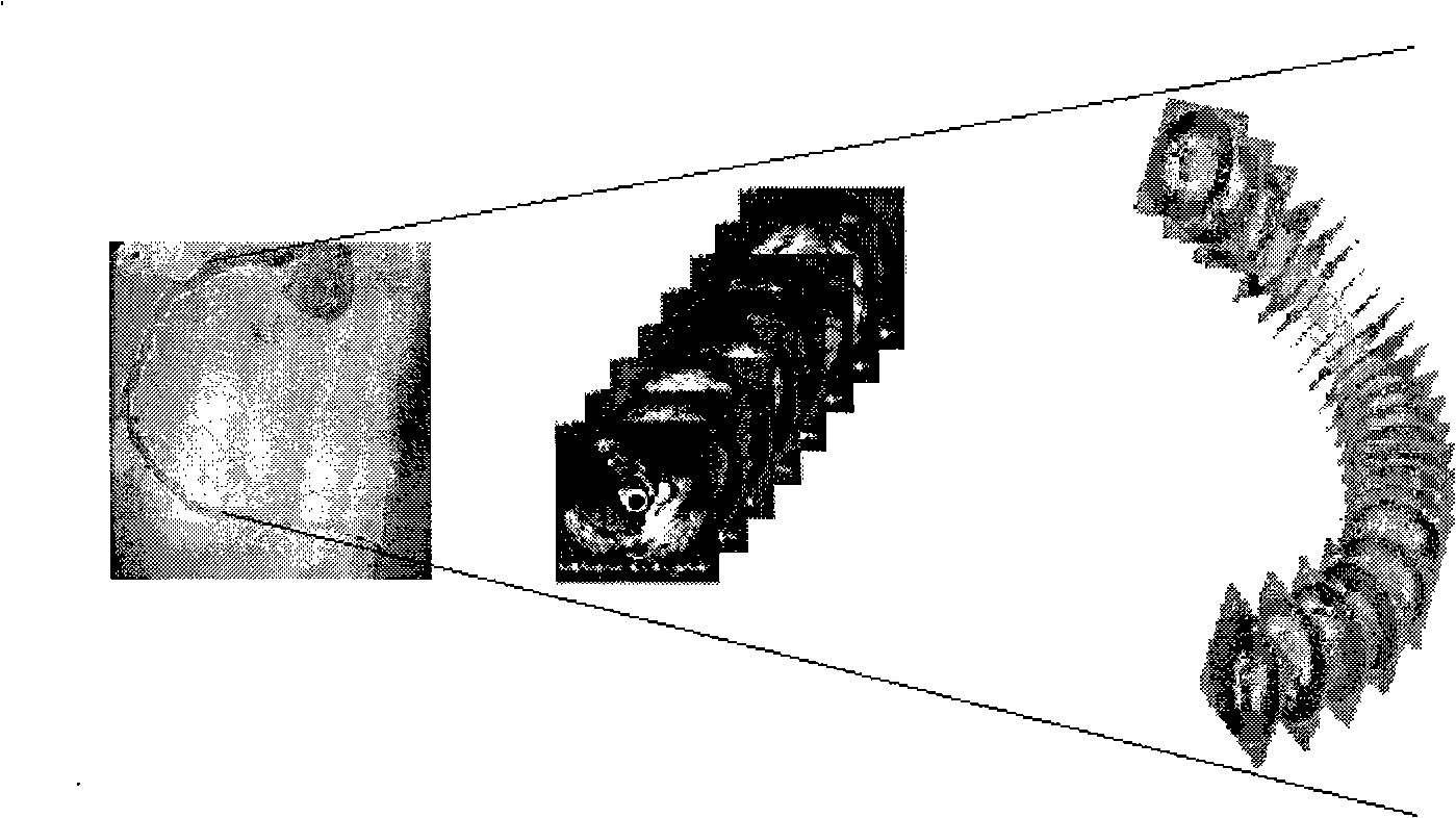
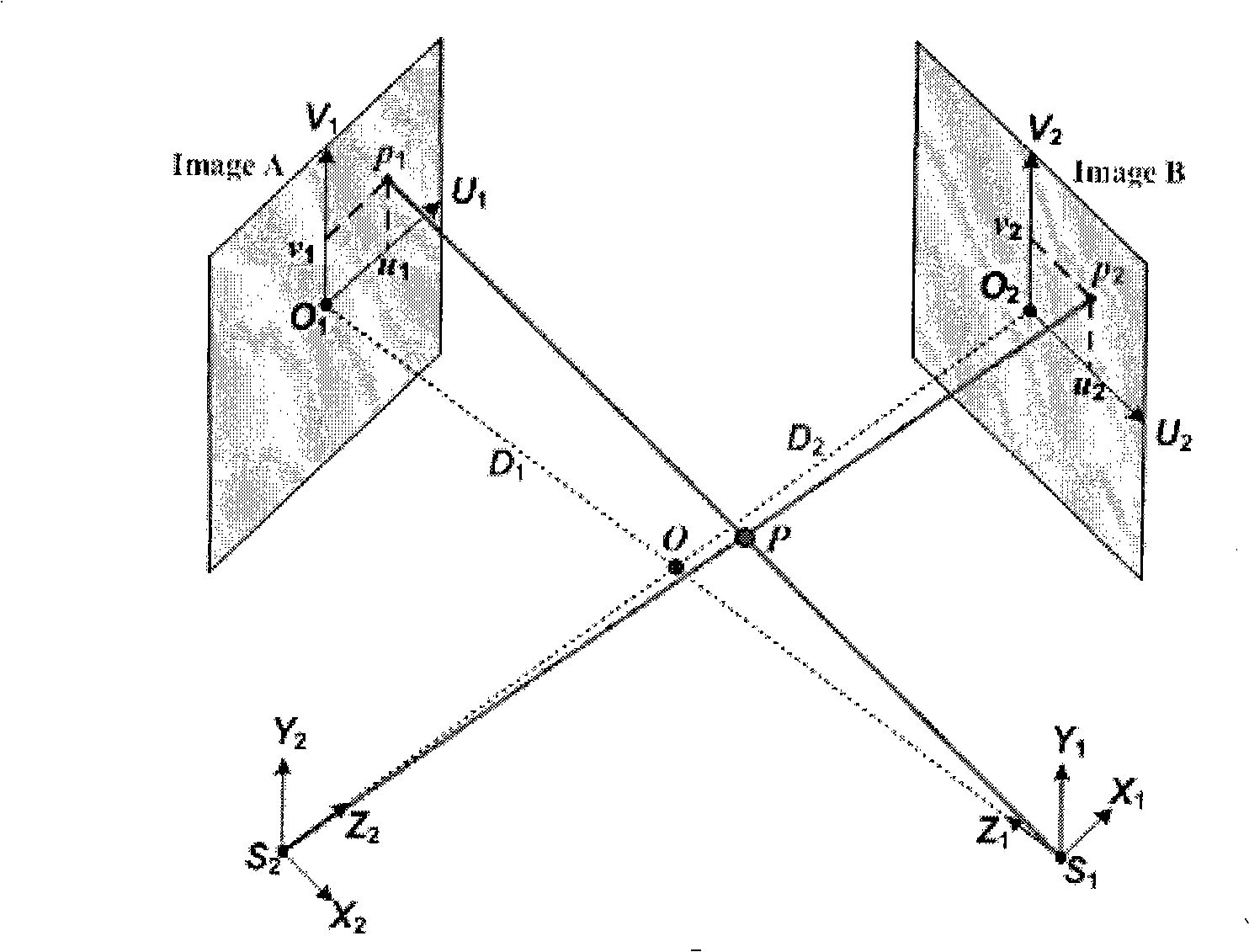
[0043] IVUS and CAG imaging were performed simultaneously (attached figure 2 ). Under the guidance of the X-ray fluoroscopy image, the guide wire with the high-frequency ultrasonic probe on the top passes through the lesion to reach the distal end of the blood vessel. Withdraw the guide wire and record the image. During the imaging process, the imaging angle and the distance from the focus of the X-ray source to the receiving screen were recorded.

[0044] For the acquisition of IVUS images, due to the periodic motion of the heart and the influence of respiration, it is difficult to obtain an image sequence corresponding to the same moment. Therefore, the present invention adopts the method of ECG (e...


PUM


Abstract

A method for reconstructing 3D blood vessels belongs to the field of medical detection technology for solving the accuracy problem in blood vessel reconstruction. The technical scheme is that the method can accurately reconstruct the anatomic structure of a blood vessel by combining cross section information of the blood vessel acquired by an IVUS image sequence and ultrasonic duct spatial geometrical information obtained by 3D reconstruction based on X-ray angiographic images. Compared with the reconstruction result obtained by using two single images, the method can completely and accurately reflect the actual shapes of blood vessels and possibly-existing plaques, thus providing more reliable basis for clinical treatment of coronary heart diseases and other angiopathies.

Description

technical field [0001] The invention relates to a blood vessel three-dimensional reconstruction method based on X-ray angiography and intravascular ultrasonic images, and belongs to the technical field of medical detection. Background technique [0002] X-ray coronary angiography (coronary angiography, CAG) examination is currently internationally recognized as the "gold standard" for the diagnosis of coronary heart disease. The specific implementation method is: use a special catheter to percutaneously puncture the femoral artery or the brachial artery of the upper arm, insert the catheter, push it to the root of the aorta under the guidance of the X-ray projection image, place it at the coronary artery, and then inject a contrast agent. Cinematography or continuous filming. Stenotic lesions caused by atherosclerosis can be found by repeated shooting from different angles several times, and the location, nature and degree of abnormal vascular anatomy can be judged based on...

Claims


Application Information

Patent Timeline
IPC IPC(8): A61B19/00A61B6/00A61B8/00G06T7/00G06T15/00A61B34/10
Inventor 孙正
Owner NORTH CHINA ELECTRIC POWER UNIV (BAODING)
Why Eureka
Try Eureka