Visible gynaecologic opto-acoustic diagnosis and treatment apparatus

A photoacoustic and gynecological technology, applied in the field of medical devices, can solve problems such as inability to diagnose, lack of automation, hidden dangers, etc., and achieve the effects of promoting blood vessel proliferation, improving nerve ending nutrition, restoring color and elasticity

Inactive Publication Date: 2009-01-14
SHANGHAI JIAO TONG UNIV
View PDF1 Cites 4 Cited by
  • Summary
  • Abstract
  • Description
  • Claims
  • Application Information

AI Technical Summary

Problems solved by technology

[0004] At present, there are following deficiencies in the prior art: (1) can only treat but not diagnose
From the perspective of the overall concept of treating gynecological diseases, it is necessary to be able to enter the treatment state immediately after diagnosis, and these problems have not been solved in the prior art; (3) Individualized treatment condition selection and control technology is relatively backward, drone dialogue measures
Especially in terms of treatment frequency, power, and time optimization selection, the best treatment mode cannot be selected according to the condition, and the degree of automation is not enough; (4) In the case of uncontrollable temperature in the ultrasonic focal region, it is impossible to quickly know the effect of the organism on the treatment process online. The information that causes the rapid response of biological tissues in the treatment area, such as temperature, pain information, behavior information, especially in the case of analgesic injection, these information can no longer be clearly transmitted to the treating doctor through the human nervous system, so it will There are some unsafe hidden dangers; (5) There is no historical record of the treatment process and results, and patients cannot observe the tissue microscopic appearance before and after treatment online; (5) The cost performance is relatively low
Such things can no longer meet the actual requirements of treating women's diseases

Method used


Image

  • Visible gynaecologic opto-acoustic diagnosis and treatment apparatus
  • Visible gynaecologic opto-acoustic diagnosis and treatment apparatus
  • Visible gynaecologic opto-acoustic diagnosis and treatment apparatus

Examples


Embodiment Construction

[0024] The embodiments of the present invention are described in detail below in conjunction with the accompanying drawings: this embodiment is implemented on the premise of the technical solution of the present invention, and detailed implementation methods and specific operating procedures are provided, but the protection scope of the present invention is not limited to the following the described embodiment.

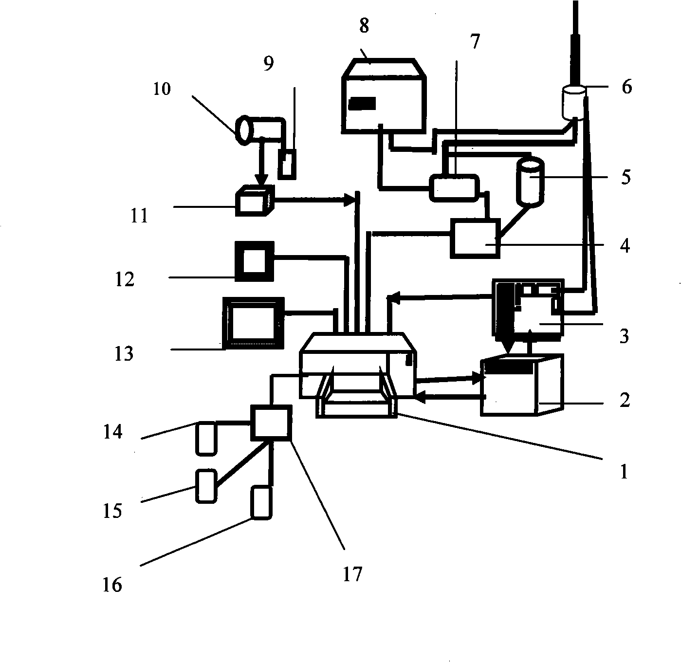
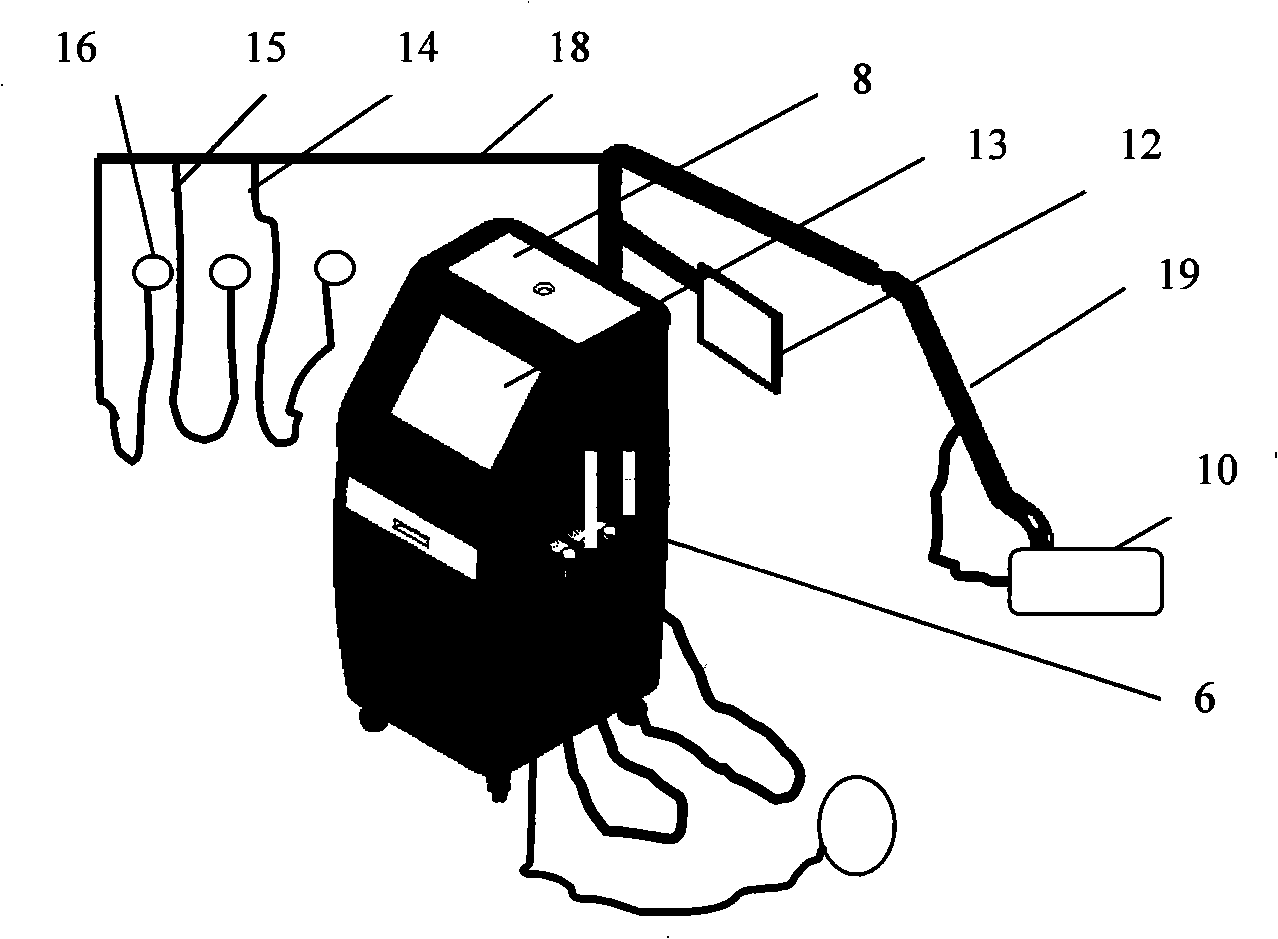
[0025] Such as figure 1 , 2 As shown, in this embodiment, it includes: a computer workstation 1, a visual observation subsystem, an ultrasonic focus therapy subsystem, and a biological pain monitoring subsystem, wherein:

[0026] The visual observation subsystem includes: a main articulated arm 9, a colposcope 10, a secondary display 12, an image acquisition card 11, and a main display 13. The colposcope 10 is fixed on one end of the main articulated arm 9, and the colposcope 10 communicates with the computer through the image acquisition card 11. The workstations 1...


PUM


Abstract

The invention relates to a visualized optical-acoustic diagnosis device used for the gynecology department, which belongs to the technical field of biomedical engineering. The device comprises a computer workstation, a visualized observation subsystem, an ultrasound focusing therapy subsystem and a biological pain monitoring subsystem, wherein, the visualized observation subsystem observe and diagnose the vaginal tissue by adopting a CCDsensor array camera system based on the optical technology, the ultrasound focusing therapy subsystem performs rapid and non-traumatic treatment to gynecological diseases by adopting the temperature field of 38 to 80 DEG C formed in the position of the subcutaneous biological tissue by the ultrasound focusing technology under the visualized condition, and the biological pain monitoring subsystem records and monitors the biological information feedback of the human body in real time under the ultrasound focusing treatment condition. By adopting the visualized optical-acoustic diagnosis device, the treatment of the gynecological diseases has higher security, the superficial layer and the adjacent tissue are not damaged, and simultaneously the visibility exists during the treatment process and the diagnosis process.

Description

technical field [0001] The invention relates to a device in the technical field of medical devices, in particular to a visualized photoacoustic diagnosis and treatment device for gynecology. Background technique [0002] As early as 1942, Lynn discovered the phenomenon of high-intensity focused ultrasound (High-intensity Focused Ultrasound). Since then, ultrasonic focusing technology has been widely used in industry, military and other fields. The application in medicine is mainly reflected in two aspects, 1, ultrasound focused imaging technology for disease diagnosis; 2, high-intensity focused ultrasound treatment of tumor technology. To this end, different types of focused ultrasonic therapy systems, ultrasonic focused therapy heads based on piezoelectric transducers, temperature monitoring technology, etc. have been invented. In 1956, Burov used high-intensity focused ultrasound (HIFU) technology for the first time to treat malignant tumors. In 1989, Umemura.S in Japan r...

Claims


Application Information

Patent Timeline
Patent Type & Authority Applications(China)
IPC IPC(8): A61B1/303A61B1/04A61N7/02
Inventor 惠春张艳丽于立立徐爱兰
Owner SHANGHAI JIAO TONG UNIV
Why Eureka
Try Eureka