Interactive coronary artery virtual angioscope implementation method

A technology of coronary arteries and implementation methods, applied in the field of medical imaging, to achieve the effect of solving motion artifacts

Inactive Publication Date: 2009-05-06
NORTH CHINA ELECTRIC POWER UNIV (BAODING)
View PDF0 Cites 43 Cited by
  • Summary
  • Abstract
  • Description
  • Claims
  • Application Information

AI Technical Summary

Problems solved by technology

Currently there is no interactive coronary virtual angioscopy system based on fusion of CAG and IVUS images that can visualize coronary arteries in endoscopic roaming mode

Method used


Image

  • Interactive coronary artery virtual angioscope implementation method
  • Interactive coronary artery virtual angioscope implementation method
  • Interactive coronary artery virtual angioscope implementation method

Examples


Embodiment Construction

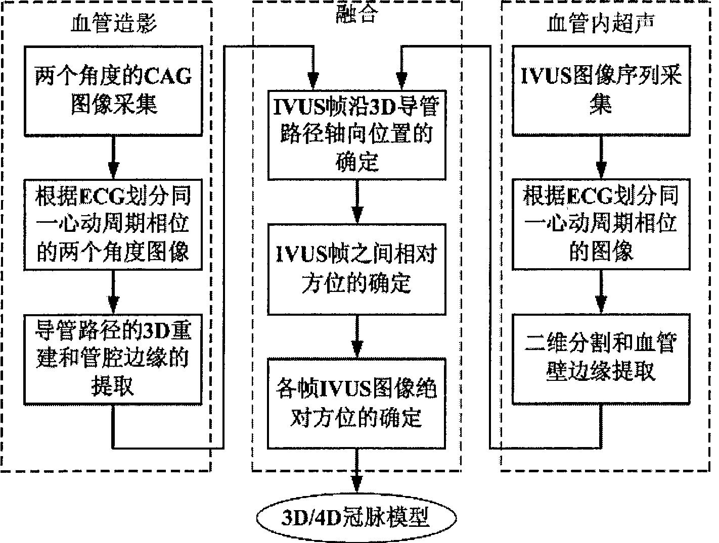
[0050] The steps of the present invention are described in detail below in conjunction with accompanying drawings:

[0051] (1) Image acquisition:

[0052] Acquisition equipment includes a C-arm single-sided X-ray angiography machine and an intravascular ultrasound imager.

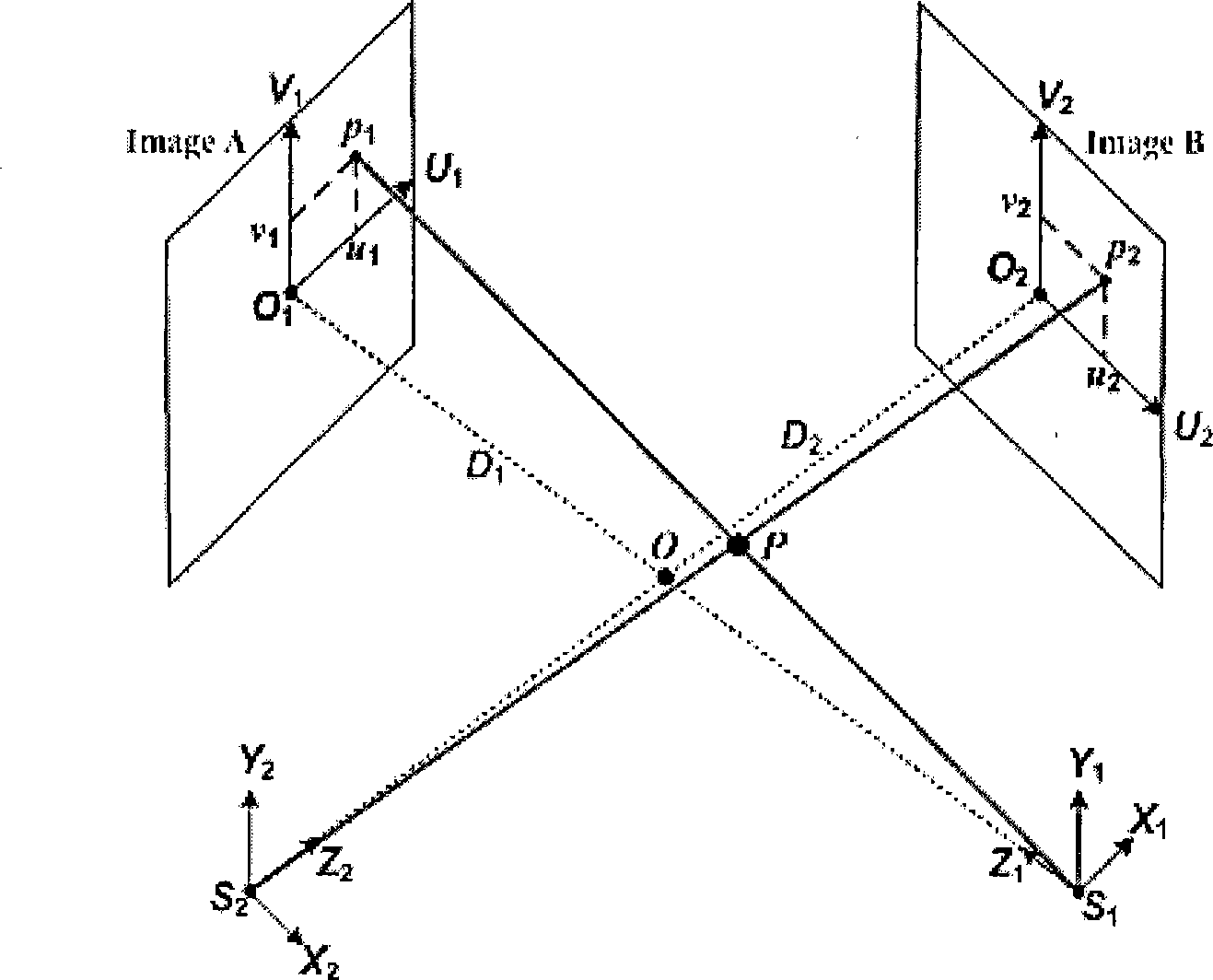
[0053] see figure 2 , IVUS and CAG imaging are performed simultaneously. Routine puncture through the right femoral artery or the brachial artery of the upper arm for selective coronary angiography. Under the guidance of the X-ray fluoroscopy image, insert the high-frequency ultrasound probe catheter to the distal end of the blood vessel. After the ultrasound probe was connected to the ultrasound imager to remove artifacts, the catheter was retracted at a constant speed and equidistant by motor control. When the probe catheter rotates 360° at 1800 r / min, real-time vascular section images of 30 frames per second are obtained continuously. The commonly used clinical method of allowing the patient to ho...


PUM


Abstract

The invention relates to a realization method for an interactive coronary artery virtual angioscope, which pertains to the technical field of medical imaging and aims at solving the visible problem of making a diagnosis and giving treatment of coronary artery. The technical proposal is that the realization method obtains the three-dimensional model of blood vessels by means of combining three-dimensional geometrical feature information of lumen by a nearly orthorhombic X-ray visualization picture of coronary artery with the data of the section of lumen by lumen endobronchial ultrasonography, then adopts virtual reality modeling language to interactively describe the model of blood vessels and realizes the visualization of coronary artery in the endoscope roaming mode. The realization method realizes interactive visit and display of the three-dimensional model of blood vessels and provides an ideal platform for the research on the development of coronary atherosclerosis pathological changes, the visible diagnosis and treatment of coronary heart diseases, the involvement in the evaluation of curative effect, and the like, and the training of medical personnel.

Description

technical field [0001] The invention relates to a method for realizing an interactive coronary virtual angioscope based on fusion of multiple imaging methods, which belongs to the technical field of medical imaging. Background technique [0002] Virtual Endoscope technology (Virtual Endoscope, VE) uses medical images as raw data, comprehensively utilizes digital image processing, computer graphics, scientific computing visualization, virtual reality and other technologies to reconstruct three-dimensional images and form virtual human tissues; Put it into the reconstructed organ cavity, and use navigation or roaming technology and pseudo-color technology to realistically simulate cavity endoscopy. [0003] Coronary Artery Angiography (CAG, Coronary Artery Angiography) and Intravascular Ultrasound (IVUS, Intravascular Ultrasound) are widely used clinically at present. The position of the ultrasound probe in the lumen and the structure of the corresponding vessel wall. CAG an...

Claims


Application Information

Patent Timeline
Patent Type & Authority Applications(China)
IPC IPC(8): A61B1/00A61B5/00G06T15/00
Inventor 孙正
Owner NORTH CHINA ELECTRIC POWER UNIV (BAODING)
Why Eureka
Try Eureka