Laser co-focusing micro-endoscope

A laser confocal and endoscopy technology, applied in the field of laser confocal microendoscopy, can solve the problem of inability to take into account small size and large scanning range, complex design of microscopic objective lens, and inability to correct the image of the endoscope system. Poor and other problems, to achieve the effect of close image resolution, no signal crosstalk, and high image contrast

Inactive Publication Date: 2009-06-10
ZHEJIANG UNIV
View PDF0 Cites 40 Cited by
  • Summary
  • Abstract
  • Description
  • Claims
  • Application Information

AI Technical Summary

Problems solved by technology

[0003] The current laser confocal microendoscope uses a fiber bundle or a single fiber to introduce laser and fluorescent signals into the body and out of the body respectively. The fiber bundle allows the confocal scanning mechanism to be placed outside the body. The endoscopic probe can be small, but the image The definition is seriously affected by the grid arrangement of the fiber bundle itself, the resolution of the confocal image is low, and the contrast of the image is difficult to improve due to the crosstalk of light from adjacent core diameters; a single fiber requires that the confocal scanning mechanism must be placed in the In the body, although the existing MEMS technology can reduce the size of the scanning mechanism, the current technology cannot take into account the small size and large scanning range; neither the optical fiber bundle nor the single optical fiber can correct the aberration of the endoscopic system
Therefore, the optical design of this type of microendoscope, especially the design of the microscopic objective lens is complex

Method used


Image

  • Laser co-focusing micro-endoscope
  • Laser co-focusing micro-endoscope
  • Laser co-focusing micro-endoscope

Examples


Embodiment Construction

[0022] The present invention will be described in detail below in conjunction with the accompanying drawings.

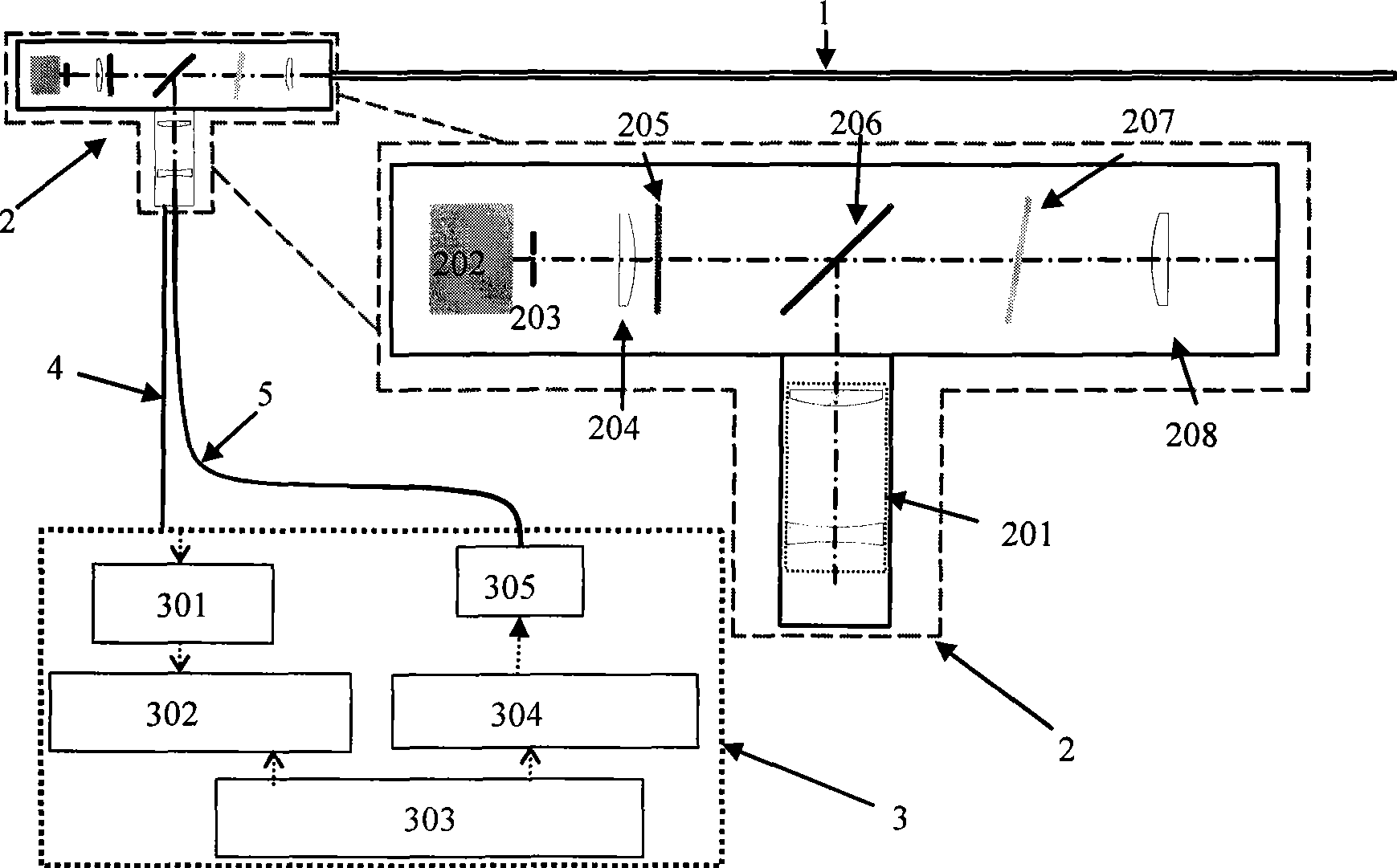
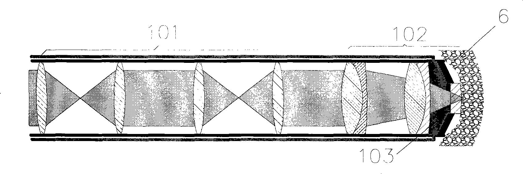
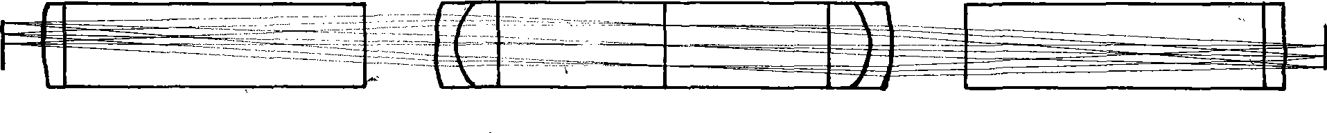
[0023] Such as figure 1 , 2 As shown, the laser confocal microendoscope of the present invention includes a microendoscope probe 1, a confocal scanning module 2, a power supply and an illumination module 3, a cable 4 and a light guide 5, and the microendoscope probe 1 includes a scope The remote image transmission system 101, the microscope objective lens 102, the scanning cavity 103, the confocal scanning module 2 includes a laser beam expander 201, a photodetector 202, a confocal diaphragm 203, a fluorescent converging lens 204, a fluorescent color filter 205, Dichroic mirror 206 , two-dimensional confocal scanner 207 , scanning objective lens 208 , power supply and illumination module 3 includes signal processing unit 301 , image workstation 302 , power supply 303 , laser driver 304 , and laser 305 .

[0024] The microendoscope probe 1 is fixed on a small confoc...


PUM


Abstract

The invention discloses a novel laser confocal microscopic endoscope with high resolution, a telescopic image transmission system is used for replacing optical fiber bundles or a single optical fiber, the laser is coupled into a tissue in vivo, a reflected light or a fluorescent signal is coupled into a detector in vitro, a fluorescent image with high resolution and contrast is obtained, meanwhile an endoscope probe diameter is reduced; a confocal scanning mechanism is arranged in vitro, the high performance confocal scanning is obtained, meanwhile the dimension of the endoscope probe can not be increased, and the confocal scanning mechanism is compatible with an existing hard tube endoscope, thereby implementing the high-performance histopathology diagnosis in vivo.

Description

technical field [0001] The invention belongs to the technical field of medical device manufacturing, in particular to a laser confocal microendoscope. Background technique [0002] Medical endoscope is an essential medical equipment for detection and diagnosis of internal lesions and minimally invasive surgery, and it is widely used in various fields of clinical medicine. Endoscopy observes internal tissues on a macro scale, identifies suspicious areas, and if necessary, clamps suspicious tissues outside the body for histopathological diagnosis, but this is a traumatic process, often accompanied by bleeding, infection, early missed diagnosis, etc. It has certain risks. Laser confocal microendoscopy can realize real-time high-resolution histopathological diagnosis of internal organs without sampling biopsy, and can use fluorescent contrast agent, which has strong specificity. It is an important tool for non-invasive diagnosis of early lesions, especially It is of great sign...

Claims


Application Information

Patent Timeline
Patent Type & Authority Applications(China)
IPC IPC(8): A61B1/07G01N21/64
Inventor 王立强石岩段会龙
Owner ZHEJIANG UNIV
Why Eureka
Try Eureka