Isolation in vitro and amplification method for adult rat heart microvascular endothelial cell

A technology of endothelial cells and microvessels, applied in artificial cell constructs, animal cells, vertebrate cells, etc.

Inactive Publication Date: 2009-06-24
FOURTH MILITARY MEDICAL UNIVERSITY
View PDF0 Cites 3 Cited by
  • Summary
  • Abstract
  • Description
  • Claims
  • Application Information

AI Technical Summary

Problems solved by technology

[0005] In related enzyme activity experiments: microvascular endothelial cells contain less Neutralendopeptidase (NEP) than large blood vessel endothelial cells, endothelial cells participate in the operation of the fibrinolyti

Method used


Image

  • Isolation in vitro and amplification method for adult rat heart microvascular endothelial cell
  • Isolation in vitro and amplification method for adult rat heart microvascular endothelial cell

Examples


Embodiment approach

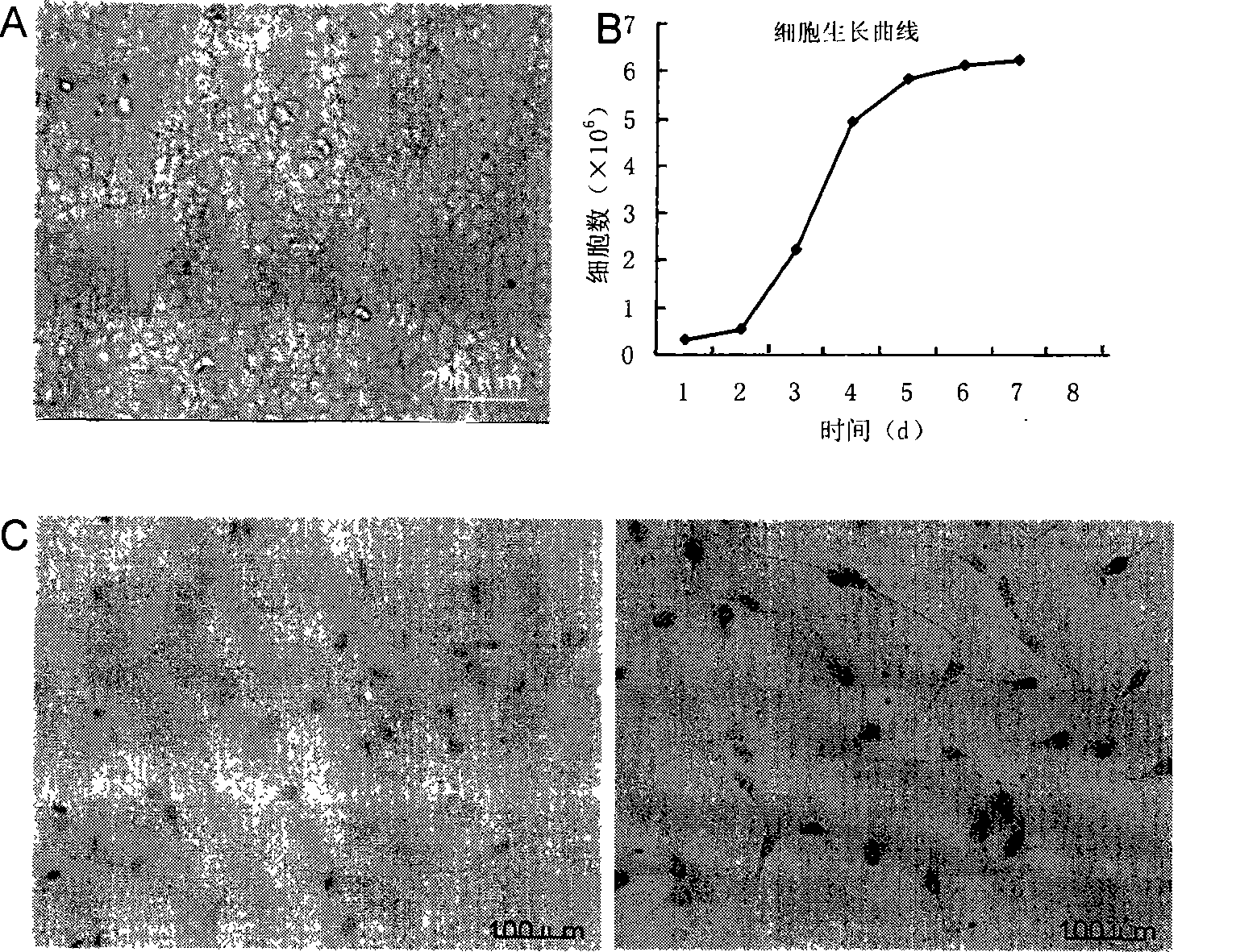
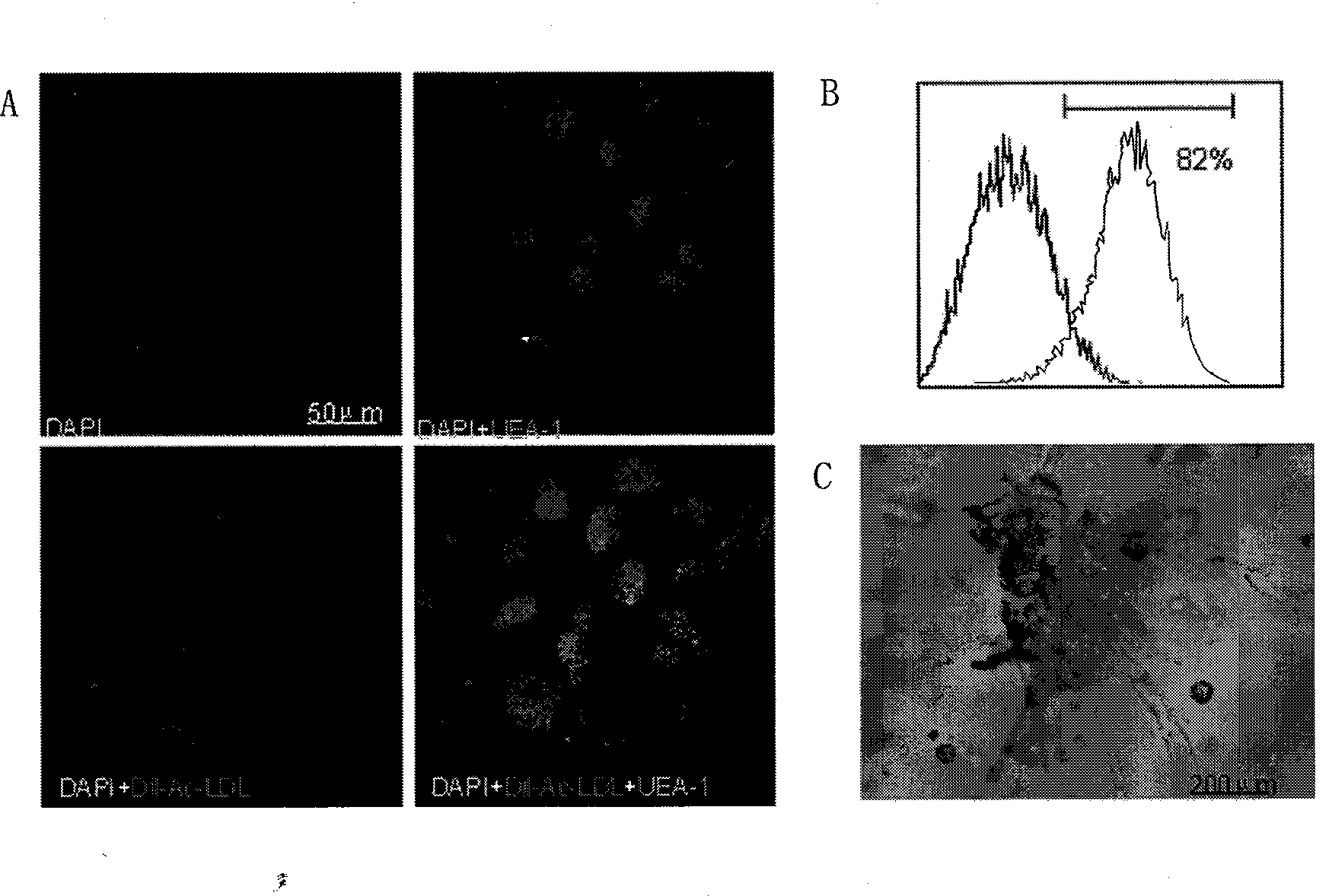
[0023] 1. Isolation and culture of endothelial cells

[0024] Get 2 (150-200g) SD rats, 3% pentobarbital sodium (30mg / Kg) intraperitoneal anesthesia, open the chest cavity, remove the heart, wash the heart cavity with PBS buffer until the flushing fluid is clear. After removing the atrium and right ventricle, cut the left ventricle along the front wall, take it out quickly after 75% alcohol inactivation for 30 seconds, and place it in PBS buffer to remove residual alcohol. Cut off the inner and outer membranes, shred the myocardial tissue, digest with 0.2% type II collagenase for 5 minutes, add 0.1% trypsin to continue the digestion for 5 minutes, and stop the digestion with DMEM medium containing 20% ​​newborn bovine serum. Collect the digestive juice and centrifuge at 1000r / min for 10min. Discard the supernatant, suspend the cells with DMEM culture medium (containing 20% ​​newborn bovine serum, penicillin 100 IU / ml, streptomycin 100 μg / ml), inoculate in a petri dish covered...


PUM


Abstract

The invention relates to a method for separating and expanding cardiac microvascular endothelial cells of adult rat vitro, which provides an ideal cell model for the correlational studies of cardiac endothelial function and a complementary source of vascular neogenesis treatment and tissue engineering cells after myocardial infarction, and lays foundation for deeply studying the clinical treatment of myocardial infarction, angiocarpy endothelial dysfunction, etc. The method adopts the technical proposal that (1) by utilizing the biological characteristics of the cardiac microvascular endothelial cells, the cardiac microvascular endothelial cells of the adult rat are separated and enriched by adopting a method of enzymic digestion, and are expanded by utilizing the self-renewing and proliferation characteristics of the endothelial cells, thus obtaining a great deal of cardiac microvascular endothelial cells; (2) the separated cells are separated and purified vitro; (3) the separated cells are purified and expanded vitro.

Description

1. Technical field: [0001] The invention relates to a method for separating and expanding adult rat cardiac microvascular endothelial cells in vitro, which is a method for utilizing the biological characteristics of rat cardiac microvascular endothelial cells, adopting collagenase rapid digestion and trypsin subculture to separate and expand rats Methods for Cardiac Microvascular Endothelial Cells. 2. Background technology: [0002] Endothelial cells of microvascular origin are different from endothelial cells of macrovascular origin, and vascular endothelial cells of different organ tissue origins also exhibit different functional properties. The endocrine function of endothelial cells is very active. Due to its special physiological location, myocardial microvascular endothelial cells are not only directly affected by blood perfusion pressure and blood flow shear stress, but also affected by repeated extrusion that other organs do not have, resulting in whether the cells i...

Claims


Application Information

Patent Timeline
IPC IPC(8): C12N5/06C12N5/071
Inventor 王海昌张荣庆曹丰魏丽萍
Owner FOURTH MILITARY MEDICAL UNIVERSITY
Why Eureka
Try Eureka