Vascular endothelial cell growth factor antagonists and uses thereof

Inactive Publication Date: 2005-03-10
GENENTECH INC
View PDF5 Cites 28 Cited by
  • Summary
  • Abstract
  • Description
  • Claims
  • Application Information

AI Technical Summary

Benefits of technology

[0012] The present invention provides antagonists of VEGF, including (a) antibodies and variants thereof which are capable of specifically binding to hVEGF, hVEGF receptor, or a complex comprising hVEGF in association with hVEGF receptor, (b) hVEGF receptor and variants thereof, and (c) hVEGF variants. The antagonists inhibit, sequester or neutralize the mitogenic, angiogenic, vascular permeability or other biological activity of hVEGF, and thus are useful for the treatment of diseases or conditions characterized by undesirable excessive neovascularization, including by way of example, tumors, and especially solid malignant tumo

Problems solved by technology

Although not fully understood, VEGF is believed to increase

Method used


Image

  • Vascular endothelial cell growth factor antagonists and uses thereof
  • Vascular endothelial cell growth factor antagonists and uses thereof
  • Vascular endothelial cell growth factor antagonists and uses thereof

Examples


example 1

Preparation of Anti-hVEGF Monoclonal Antibodies

[0109] To obtain hVEGF conjugated to keyhole limpet hemocyanin (KLH) for immunization, recombinant hVEGF (165 amino acids), Leung, et al., Science 246:1306 (1989), was mixed with KLH at a 4:1 ratio in the presence of 0.05% glutaraldehyde and the mixture was incubated at room temperature for 3 hours with gentle stirring. The mixture then was dialyzed against phosphate buffered saline (PBS) at 4° C. overnight.

[0110] Balb / c mice were immunized four times every two weeks by intraperitoneal injections with 5 μg of hVEGF conjugated to 20 μg of KLH, and were boosted with the same dose of hVEGF conjugated to KLH four days prior to cell fusion.

[0111] Spleen cells from the immunized mice were fused with P3X63Ag8U.1 myeloma cells, Yelton, et al., Curr. Top. Microbiol. Immunol. 81:1 (1978), using 35% polyethylene glycol (PEG) as described. Yarmush, et al., Proc. Nat. Acad. Sci. 77:2899 (1980). Hybridomas were selected in HAT medium.

[0112] Super...

example 2

Characterization of Anti-hVEGF Monoclonal Antibodies

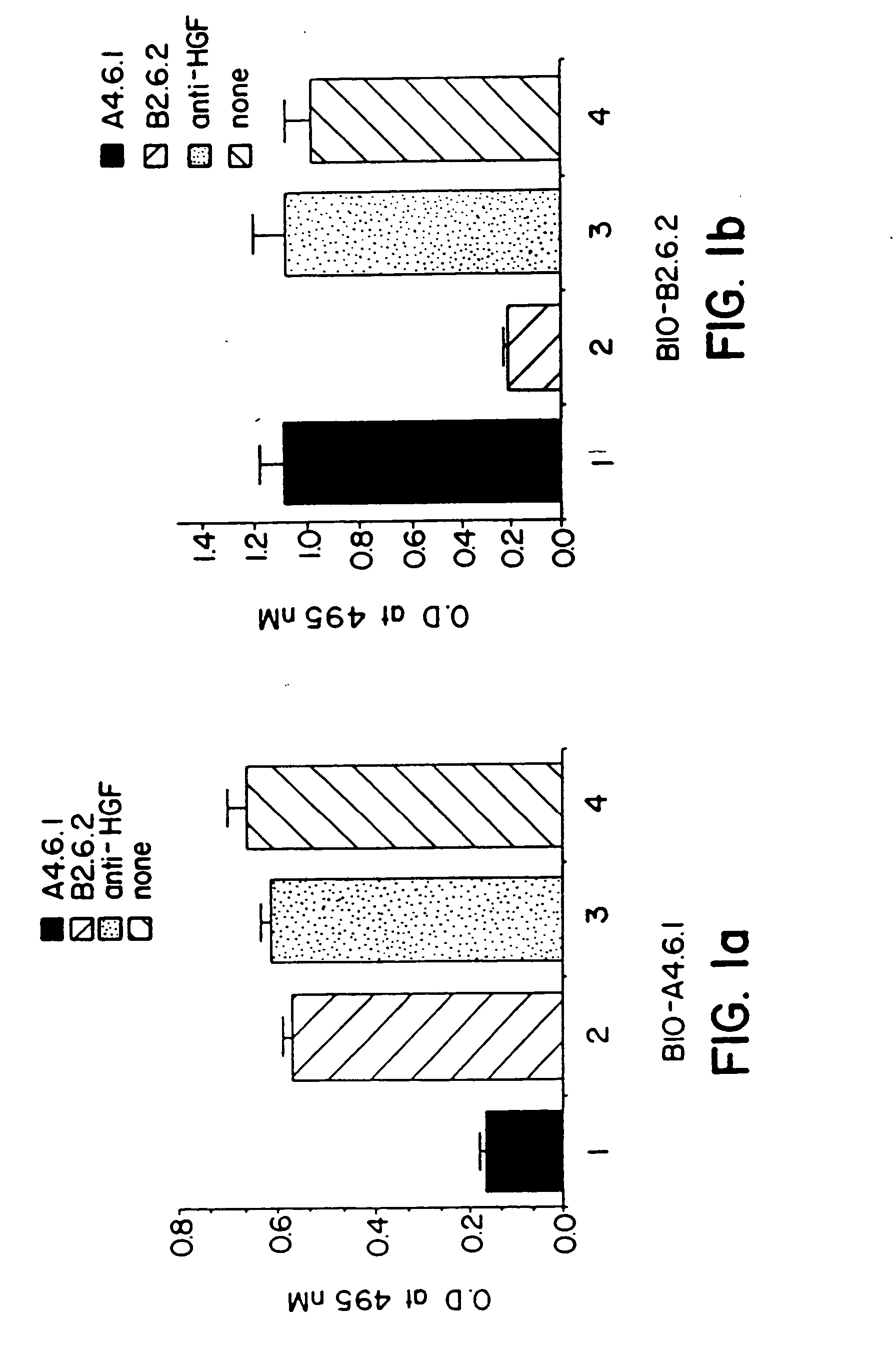
[0113] A. Antigen Specificity

[0114] The binding specificities of the anti-hVEGF monoclonal antibodies produced by the A4.6.1 and B2.6.2 hybridomas were determined by ELISA. The monoclonal antibodies were added to the wells of microtiter plates that previously had been coated with hVEGF, FGF, HGF, or epidermal growth factor (EGF). Bound antibody was detected with peroxidase conjugated goat anti-mouse IgG immunoglobulins. The results of those assays confirmed that the monoclonal antibodies produced by the A4.6.1 and B2.6.2 hybridomas bind to hVEGF, but not detectably to those other protein growth factors.

[0115] B. Epitope Mapping

[0116] A competitive binding ELISA was used to determine whether the monoclonal antibodies produced by the A4.6.1 and B2.6.2 hybridomas bind to the same or different epitopes (sites) within hVEGF. Kim, et al., Infect. Immun. 57:944 (1989). Individual unlabeled anti-hVEGF monoclonal antibodies (A4.6.1 or B...

example 3

Preparation of VEGF Receptor—IQG Fusion Proteins

A.

[0137] The nucleotide and amino acid coding sequences of the flt hVEGF receptor are disclosed in Shibuya, et al., Oncogene 5:519-524 (1990). The coding sequence of the entire extracellular domain of the flt hVEGF receptor was fused to the coding sequence of human IgG1 heavy chain in a two-step process.

[0138] Site-directed mutagenesis was used to introduce a BstBI restriction into DNA encoding fit at a site 5′ to the codon for amino acid 759 of fit, and to convert the unique BstEII restriction site in plasmid pBSSK-FC, Bennett, et al., J. Biol. Chem. 266:23060-23067 (1991), to a BstBI site. The modified plasmid was digested with EcoRI and BstBI and the resulting large fragment of plasmid DNA was ligated together with an EcoRI-BstBI fragment of the fit DNA encoding the extracellular domain (amino acids 1-758) of the flt hVEGF receptor.

[0139] The resulting construct was digested with ClaI and NotI to generate an approximately 3.3 kb...


PUM

PropertyMeasurementUnit
Densityaaaaaaaaaa
Volumeaaaaaaaaaa

Abstract

The present invention provides vascular endothelial cell growth factor (VEGF) antagonists and methods of using VEGF antagonists. VEGF antagonists contemplated by the invention include VEGF antibodies and VEGF receptor fusion proteins. Methods of treating edema and stroke using VEGF antagonists are also provided.

Description

[0001] This is a non-provisional application filed pursuant to 37 CFR 1.53(b). FIELD OF THE INVENTION [0002] The present invention relates to vascular endothelial cell growth factor (VEGF) antagonists, to therapeutic compositions comprising the antagonists, and to methods of use of the antagonists for diagnostic and therapeutic purposes. In particular, the present invention relates to methods of treatment of stroke or edema using VEGF antagonists. BACKGROUND OF THE INVENTION [0003] The two major cellular components of the vasculature are the endothelial and smooth muscle cells. The endothelial cells form the lining of the inner surface of all blood vessels, and constitute a nonthrombogenic interface between blood and tissue. In addition, endothelial cells are an important component for the development of new capillaries and blood vessels. Thus, endothelial cells proliferate during the angiogenesis, or neovascularization, associated with tumor growth and metastasis, as well as a vari...

Claims


Application Information

Patent Timeline
IPC IPC(8): A61K38/00C12N15/09A61K39/395A61K45/00A61P3/10A61P7/10A61P9/10A61P11/00A61P17/06A61P19/02A61P27/02A61P27/06A61P29/00A61P35/00A61P43/00C07K14/71C07K16/22C07K16/24C12N9/96
CPCA61K2039/505C07K2317/76C07K16/22C07K16/24C07K2316/96C07K2317/73C07K2317/92C07K2319/30C12N9/96A61K38/00A61K39/3955A61K39/39533A61K38/179C07K16/18A61N5/10A61K45/06C07K14/71A61P11/00A61P17/06A61P19/02A61P25/00A61P27/02A61P27/06A61P29/00A61P35/00A61P43/00A61P7/00A61P7/10A61P9/00A61P9/10A61P3/10Y02A50/30
Inventor VAN BRUGGEN, NICHOLASFERRARA, NAPOLEONE
Owner GENENTECH INC
Why Eureka
Try Eureka