Functional pigmented skin equivalent

An equivalent, skin technology, applied in the direction of epidermal cells/skin cells, microorganisms, animal cells, etc., can solve the problems that models cannot be displayed, melanin is not observed, and lack of control of epidermal cells

Active Publication Date: 2009-09-23
LOREAL SA
View PDF2 Cites 17 Cited by
  • Summary
  • Abstract
  • Description
  • Claims
  • Application Information

AI Technical Summary

Problems solved by technology

Among the disadvantages of this technique, the lack of control of epidermal cells needs to be mentioned, as well as the fact that only one skin reconstruction is possible with biopsies: therefore no experiments are available for compa

Method used


Image

  • Functional pigmented skin equivalent
  • Functional pigmented skin equivalent
  • Functional pigmented skin equivalent

Examples


Embodiment 1

[0162] Example 1: Preparation of skin equivalents

[0163] Preparation of lattices (D-4)

[0164] Dermis equivalents were prepared using bovine type I collagen and human fibroblasts derived from the dermis.

[0165] Bovine type I collagen and human Fibroblasts prepare dermal equivalents. The Petri dish was pre-coated with MEM containing 10% serum.

[0166] A solution of laminin (3% / final volume), type IV collagen (1.5% / final volume), and nestin (0.35% / final volume) was added to the mixture before pouring into the collagen preparation containing cells, vigorously Stir. Place the suspension in an incubator (37 °C-5% CO 2 ) to enable the grid to shrink.

[0167] Inoculation of keratinocytes and melanocytes (D0)

[0168] After retraction of the lattice, keratinocytes and melanocytes were seeded on dermal equivalents.

[0169] For this, cells were expanded for 7 days in their respective growth media before seeding:

[0170] Melanocytes were expanded in the medium for...

Embodiment 2

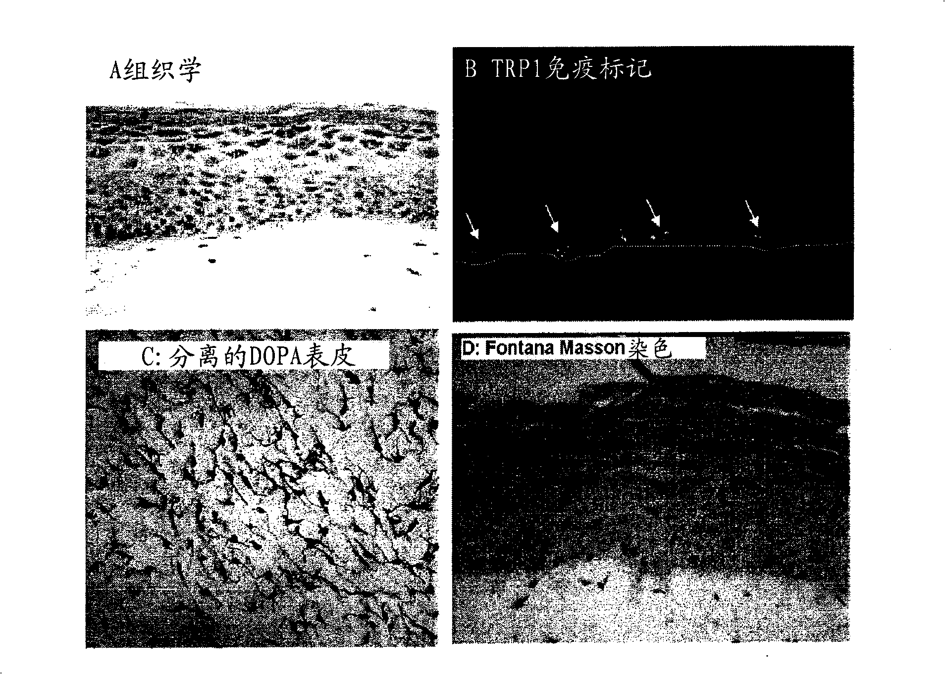
[0180] Example 2: Human Reconstituted Pigmented Skin Containing 3 Cell Types: Melanocytes, Keratinocytes and fibroblasts ( figure 1 )

[0181] After 7 days of emergence, the skin exhibited a histology and three-dimensional structure similar to human skin.

[0182] It has a stratified and differentiated epidermis composed of various cell layers (basal, spinous-cell, granular) and stratum corneum.

[0183] The dermis contains viable fibroblasts embedded in a collagen matrix ( figure 1 A).

[0184] Melanocytes are integrated into the epidermis and display a morphology and density similar to normal skin ( figure 1 C, DOPA response on isolated epidermis). They are clearly localized in the basal layer ( figure 1B, Immunolabeling of TRP1, the dotted white line indicates the dermal-epidermal junction), and the production of melanosomes and melanin, which are transferred to nearby keratinocytes ( figure 1 D, using Fontana-Masson to stain melanin).

Embodiment 3

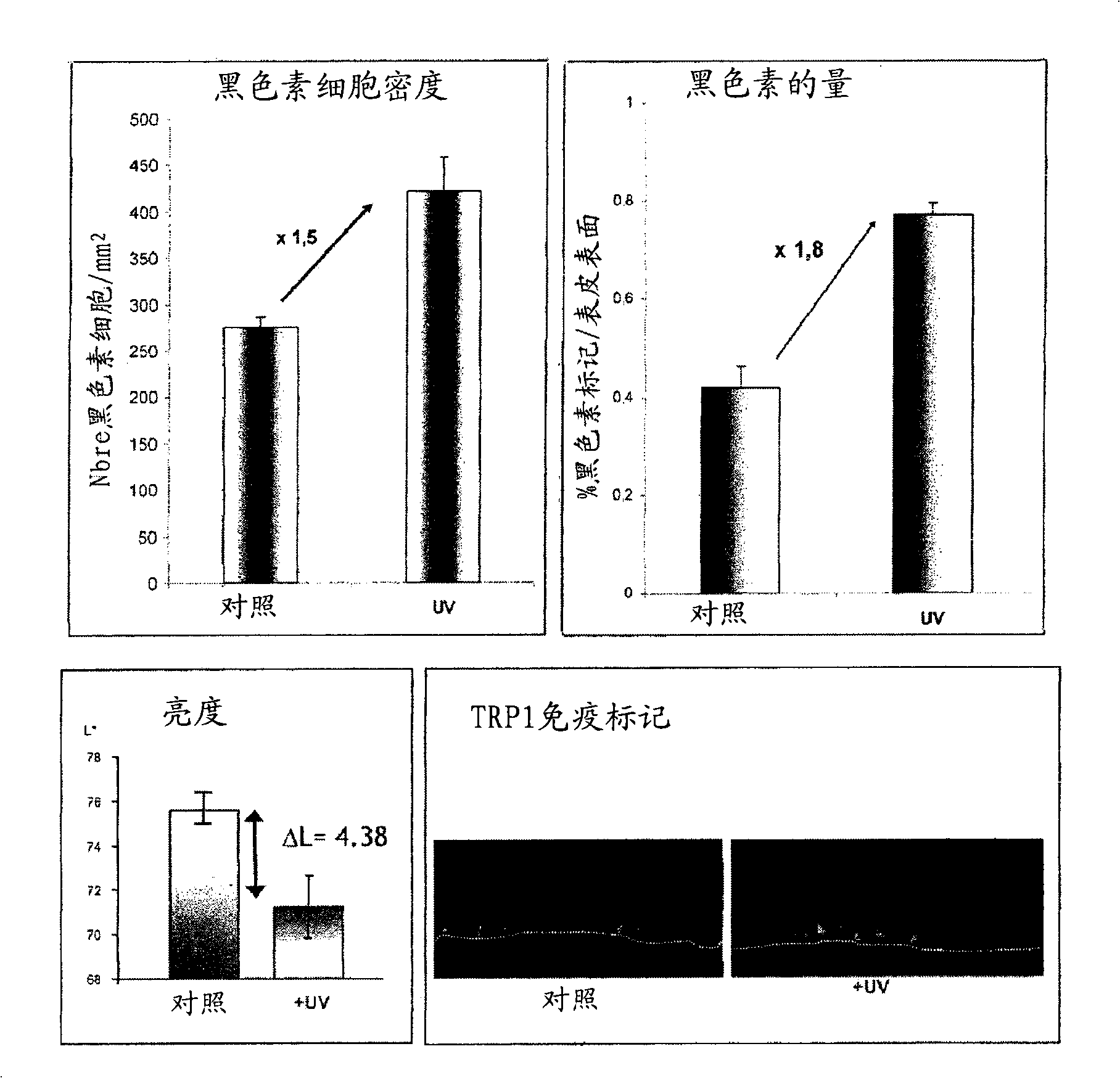
[0185] Example 3: Human Reconstituted Pigmented Skin with Constitutive Pigment Shape Associated with Melanocytes to make( figure 2 )

[0186] The model was able to integrate various melanocyte lines from the least pigmented to the most highly pigmented (sound model), the phenotype of the more or less pigmented reconstructed skin corresponded to the melanocyte type used ( Physical maintenance): In fact, the more pigment the melanocyte line has (M03-M504), the darker the macroscopic color of the skin, and the greater the amount of melanin visible on the slice. This macroscopic pigmentation is reflected by a decrease in brightness ( figure 2 ,brightness).


PUM


Abstract

The present invention relates to a functional pigmented skin equivalent. The invention relates to an in vivo skin equivalent which is characterized by comprising at least one epidermis equivalent and at least one dermis equivalent. The in vivo skin equivalent further comprises melanocytes constitutively producing melanin and fibroblasts. The invention also relates to a method for preparing the skin equivalent.

Description

technical field [0001] The present invention relates to reconstructed skin with constitutive pigmentation, and their use in methods of assessing the phenomenon of skin pigmentation; thus, the present invention relates to methods of assessing agents capable of modulating such pigmentation, whether said pigmentation is constitutive is still induced. The invention also relates to methods for preparing such skin models. Background technique [0002] Skin color is mainly due to the presence of a pigment, melanin, in the epidermis. Melanin is synthesized by specialized dendritic cells, melanocytes, located in the basal layer of the epidermis. Melanogenogenesis occurs in organelles, and melanin-loaded melanosomes are transferred to nearby epidermal cells, keratinocytes, through dendrites. [0003] Pigmentation can be regulated by physical factors, such as UV rays, or chemical factors, such as depigmenting or propigmenting products, and / or during physiological or pathological mod...

Claims


Application Information

Patent Timeline
IPC IPC(8): C12N5/08C12Q1/02C12N5/071
CPCC12N2502/094C12N2501/117C12N2502/1323C12N2501/11C12N5/0698C12N2501/39C12N2533/50C12N2533/52C12N2533/54
Inventor C·杜瓦尔F·伯纳德
Owner LOREAL SA
Why Eureka
Try Eureka