Method for screening atherosclerosis related miRNA

A technology for atherosclerosis and screening methods, applied in the direction of biochemical equipment and methods, microbial measurement/testing, etc., to achieve the effect of perfecting the formation mechanism

Inactive Publication Date: 2010-01-13
XIN HUA HOSPITAL AFFILIATED TO SHANGHAI JIAO TONG UNIV SCHOOL OF MEDICINE
View PDF0 Cites 7 Cited by
  • Summary
  • Abstract
  • Description
  • Claims
  • Application Information

AI Technical Summary

Problems solved by technology

However, this study used LPS as a stimulating factor to induce inflammation, and THP-1 cell line was used as the research object, which simulated only the natural immune response of m

Method used


Image

  • Method for screening atherosclerosis related miRNA
  • Method for screening atherosclerosis related miRNA
  • Method for screening atherosclerosis related miRNA

Examples


Embodiment 1

[0018] Example 1 Isolation of Human Peripheral Blood Mononuclear Cells and Their Phenotypic Characterization After Different Times of Ox-LDL Inflammatory Stimulation

[0019] Heparin anticoagulated venous blood was taken from healthy adult volunteers, mononuclear cells were separated by density gradient centrifugation and adherence method, and monocytes were incubated together with ox-LDL (30mg / L) for 6 to 12 hours.

Embodiment 2

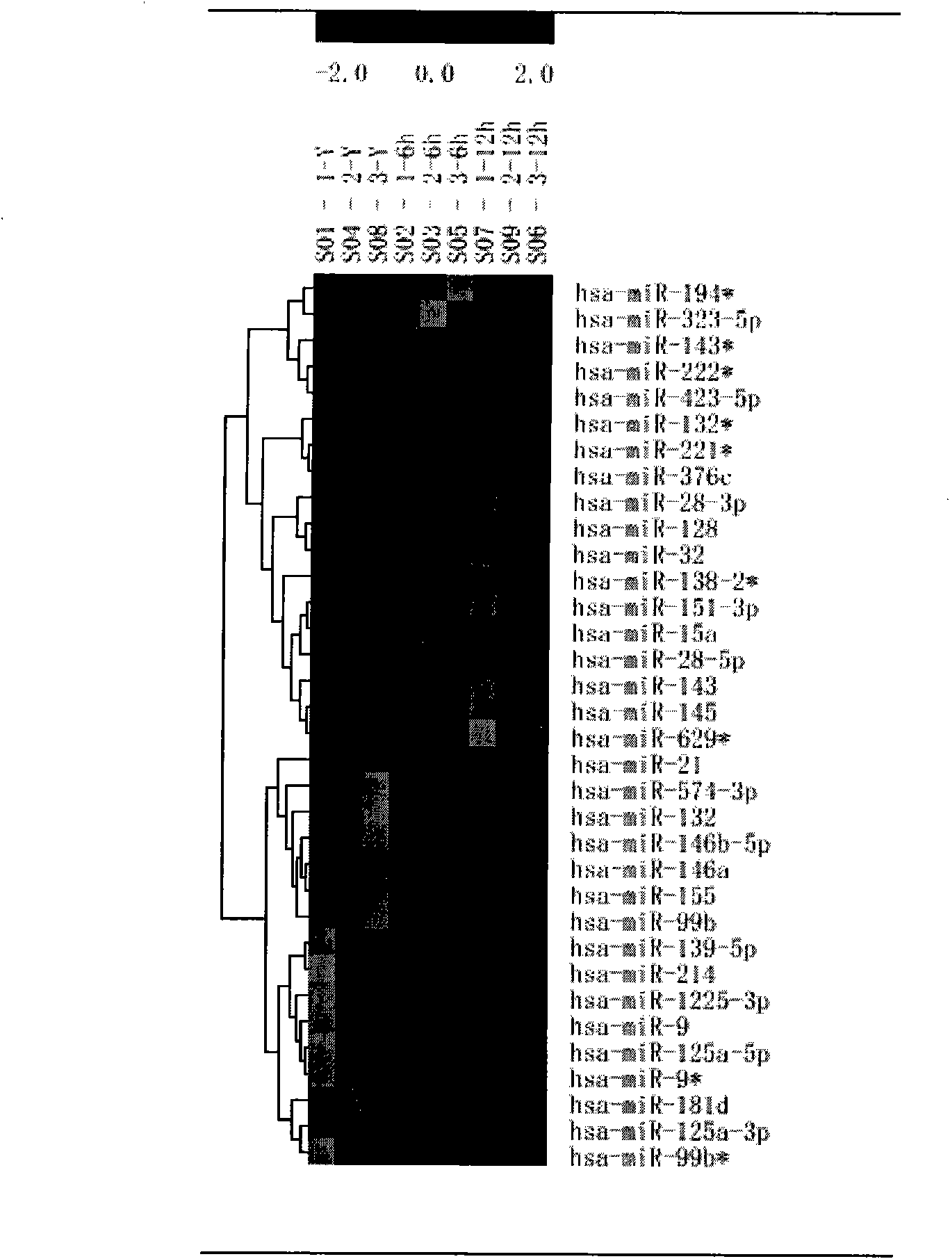
[0020] Example 2 Divided into monocyte group and ox-LDL inflammatory stimulation group at different time (6 hours, 12 hours), and Microarray analysis technology was used to detect the expression level of miRNAs in each group

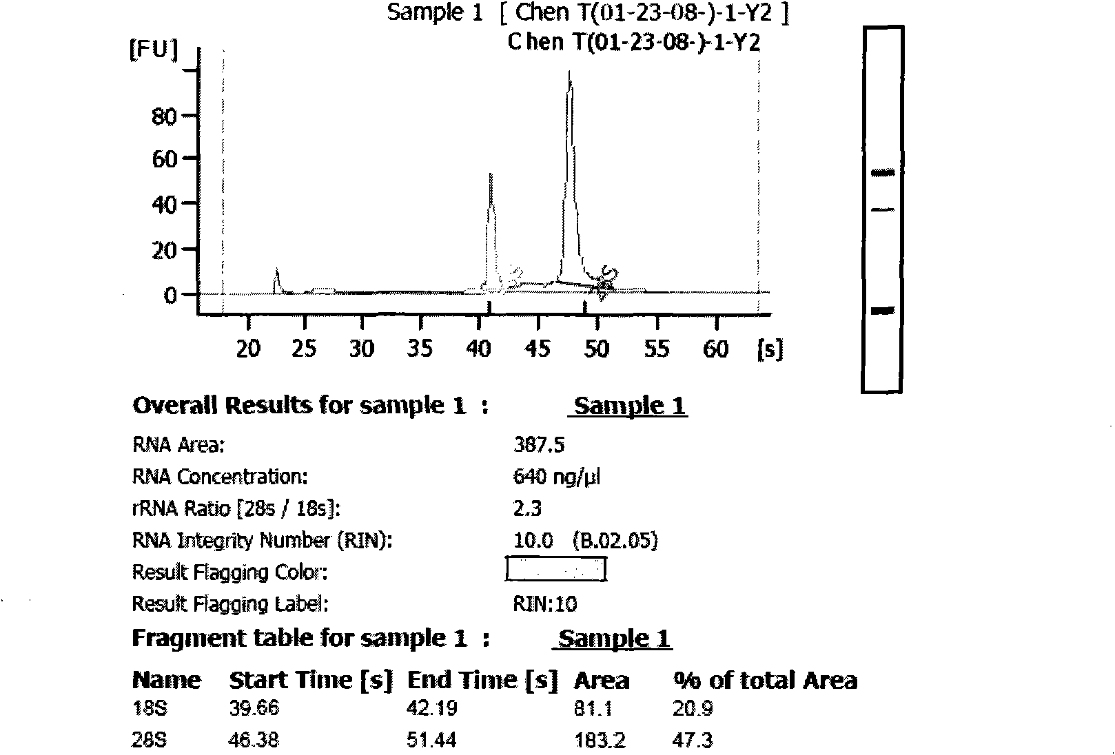
[0021] The Qiagen kit extracted more than 5 μg of total RNA samples, and separated them through YM-100 (Millipore) microcentrifuge filter columns to obtain small RNAs with fragments less than 300 nt. please see figure 1 As shown, the obvious 5S, 18S, and 28S electrophoresis bands can be seen from the figure, which proves that the small RNA is not lost.

[0022] Poly(A) polymerase adds a poly(A) tail to the 3′ end of the isolated small RNA, and then an oligonucleotide tag is ligated to the poly(A) tail for subsequent fluorescent labeling ( Cy3 and Cy5).

[0023] The labeled miRNAs were hybridized with the miRNAs chip: the hybridization reaction was performed overnight on the μParaflo microfluidic chip (Actic Technologies) by a microcirculation pump ...

Embodiment 3

[0024] Example 3 Detection confirms differential expression of miRNAs and predicts possible target mRNAs of corresponding miRNAs

[0025] The 5 miRNAs with the most significant difference between 6 and 12 hours of ox-LDL inflammatory stimulation screened by the chip were verified by fluorescent quantitative PCR on the 7000 reaction instrument with tagman probe (Applied Biosystems, Foster City, CA), and first used MuLV ( Multiscribe) reverse transcriptase and specific primers for reverse transcription, prepare a 15-μL reaction system according to the instructions, set cycle parameters at 16°C for 30 minutes, 42°C for 30 minutes, 85°C for 5 minutes, and stop temperature at 4°C. Then use RT products, Taqman 2X General PCR Master Mix and 5X MicroRNA Assay Mix containing primers and specific probes to prepare a 20-μL reaction system for amplification, set cycle parameters at 95°C for 10 minutes, 95°C for 15 seconds for denaturation and annealing amplification at 60°C for 60 seco...


PUM


Abstract

The invention discloses a method for screening atherosclerosis related miRNA, which comprises the following steps: selecting ox-LDL (30mu g/ml) as a stimulating factor to stimulate human primary monocyte and induce the human primary monocyte to be differentiated into macrophage and foam cell, detecting the difference of cellular miRNAs expression in vitro by combining microarray analysis technology, screening the miRNAs which possibly participate in atherosclerosis reaction, and then using tagman probe fluorescent quantitative PCR verification and using a database and a computer software to analyze and forecast a possible target mRNA corresponding to the screened miRNAs; therefore, the method furthest screens the miRNA close to an atherosclerosis specific inflammation reaction pathological state, further completes a forming mechanism of atherosclerosis, and develops a new treatment target and a method for preventing and treating the atherosclerosis, in particular acute coronary events.

Description

technical field [0001] The invention relates to the field of treatment of atherosclerotic diseases, in particular to a method for screening miRNAs related to atherosclerotic inflammation. Background technique [0002] Although the pathophysiological mechanism of atherosclerosis has not been fully clarified, the theory of inflammatory response is generally accepted at present. Inflammation is a key factor in the development of atherosclerosis. Studies at home and abroad have shown that there are many inflammatory factors involved in the pathological process of atherosclerosis, and the key step is that monocytes migrate into the subintimal membrane and are stimulated by local oxidized low-density lipoprotein (ox-LDL) to be activated and differentiate into macrophages. Phage cells, macrophages that have phagocytosed excess ox-LDL, differentiate into foam cells, which are not only the main component of the atherosclerotic lipid core, but also produce and release matrix metallop...

Claims


Application Information

Patent Timeline
IPC IPC(8): C12Q1/68
Inventor 王长谦陈婷黄周青王连升汪月
Owner XIN HUA HOSPITAL AFFILIATED TO SHANGHAI JIAO TONG UNIV SCHOOL OF MEDICINE
Why Eureka
Try Eureka