
Positron emission tomography (PET) and computed tomography (CT) cross-modality medical image fusion method based on wavelet transform

A fusion technology of CT images and different machines, applied in the field of image fusion, can solve problems such as imbalance, waste of resources, and insufficient equipment, and achieve excellent results, reasonable cost, and rapid results

Inactive Publication Date: 2011-03-23
徐子海 +1
View PDF0 Cites 23 Cited by
  • Summary
  • Abstract
  • Description
  • Claims
  • Application Information

AI Technical Summary

Problems solved by technology

There are many problems in the actual clinical application of the integration of the same machine: First, the fixed cost of the all-in-one machine is large, the cost recovery is difficult, and there is a serious waste of resources, and due to the economic situation and consumption level, it is limited to the nationwide equipment Insufficient and unbalanced quantity limit the clinical application and development of the same machine fusion. Simply put, the same machine equipment is very expensive. In comparison, the heterogeneous fusion is cheap and has a broad space for clinical application. ;Secondly, because the clinical needs are usually more complicated, it is impossible to combine all the required machines. If two images of the same mode are registered for the same patient at different times to measure the curative effect, software has to be used for registration. Fusion, it can be seen that software registration fusion, that is, fusion of different machines, is very necessary

Method used


Image

  • Positron emission tomography (PET) and computed tomography (CT) cross-modality medical image fusion method based on wavelet transform
  • Positron emission tomography (PET) and computed tomography (CT) cross-modality medical image fusion method based on wavelet transform
  • Positron emission tomography (PET) and computed tomography (CT) cross-modality medical image fusion method based on wavelet transform

Examples


Embodiment Construction

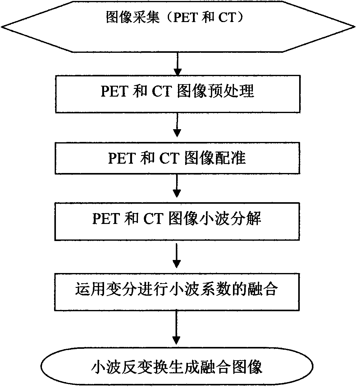
[0024] Taking the fusion of the PET image and CT image of the chest of a patient with lung cancer as an example below, the working steps of the present invention are described in detail, as figure 1 Shown:

[0025] 1. Firstly, we use the body scanning outer reference frame designed by us for PET and CT scanning respectively, and collect PET images and CT images of lung cancer patients. After our improved design of the body and head and neck scanning outer reference frame, Z The font-shaped hollow aluminum tube is inlaid in plexiglass, and FDG and copper sulfate are used as contrast agents for imaging in PET and CT scans to generate PET images and CT images with marked points. The CT image is a floating image, and the PET image is used as a reference image.

[0026] 2. The original format of the collected images is DICOM format. Use DICOM2 software to convert them into BMP format respectively for the next step of processing. The data matrix of PET images is 128×128, and that o...


PUM


Abstract

The invention discloses a discloses a positron emission tomography (PET) and computed tomography (CT) cross-modality medical image fusion method based on wavelet transform, comprising the following steps shown in the figure 1: fixing a patient in a locating frame; acquiring a PET image according to the frame and simultaneously utilizing the locating frame to acquire a CT image; respectively extracting the PET image and the CT image; registering the extracted PET image and the extracted CT image according to locating points obtained in the locating frame; carrying out wavelet decomposition and variation solving fusion on the rectified PET image and the registered PET image and the registered CT image; and finally carrying out wavelet inverse transformation on the PET image and the CT image which are subject to fusion to generate a fused image. The invention has the advantages that rectification is carried out on the acquired PET image and the acquired CT image by an external label point-based method, and a variation method is innovatively proposed to be applied to a wavelet region, thus realizing the accurate fusion of PET and CT cross-modality medical image; and the method is simple in operation and accurate in results, and has important application value and economic benefits in clinical applications.

Description

technical field [0001] The invention relates to an image fusion method, in particular to a different-machine fusion method of PET images and CT images. Background technique [0002] Radiation therapy technology has gone through several stages of development: general radiotherapy, stereotactic radiosurgery (SRS), three-dimensional conformal radiation therapy (3DCRT), intensity-modulated radiation therapy (IMRT) and image-guided radiation therapy (IGRT). In the era of precise radiotherapy, precise radiotherapy can minimize the irradiation of normal tissues around the tumor, greatly increase the irradiation dose of the tumor, and improve the local control rate of the tumor. [0003] Tumor radiation therapy is a major field of medical image fusion applications, and accurate target delineation is the basis for accurate treatment. With the development of molecular imaging, PET, that is, positron emission tomography, can display the biological characteristics of tumors, and is inc...

Claims


Application Information

Patent Timeline
IPC IPC(8): A61B6/03G06T5/00
Inventor 徐子海陈超敏童蕾周凌宏廖福锡胡赟刘凌湘
Owner 徐子海
Learn More