Method for isolating cell free apoptotic or fetal nucleic acids

A cell nucleic acid and nucleic acid technology, applied in the fields of separation of fetal nucleic acid, separation of apoptosis or fetal nucleic acid, identification of fetal genetic composition, and can solve the problem of lack of prenatal examination or screening of pregnant women

Inactive Publication Date: 2011-03-30
NOVARTIS AG
View PDF3 Cites 3 Cited by
  • Summary
  • Abstract
  • Description
  • Claims
  • Application Information

AI Technical Summary

Problems solved by technology

However, the lack of adequate or relatively safe prenatal testing or screening for most pregnant women has resulted in approximately 80% of babies born to women with Down syndrome being under the age of 35

Method used


Image

  • Method for isolating cell free apoptotic or fetal nucleic acids
  • Method for isolating cell free apoptotic or fetal nucleic acids
  • Method for isolating cell free apoptotic or fetal nucleic acids

Examples


Embodiment 1

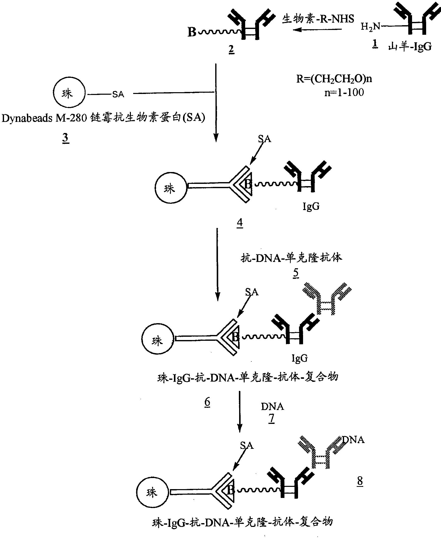
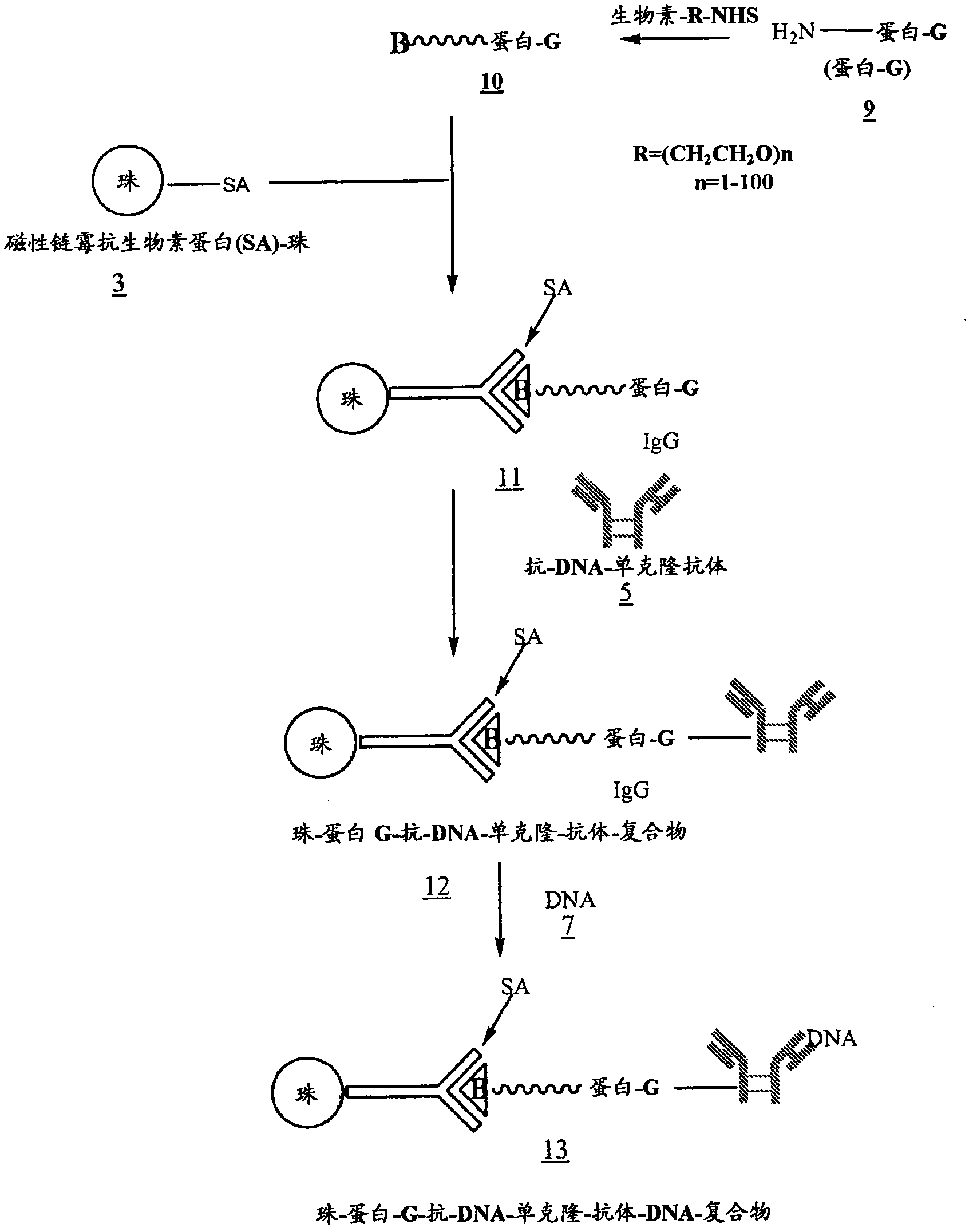
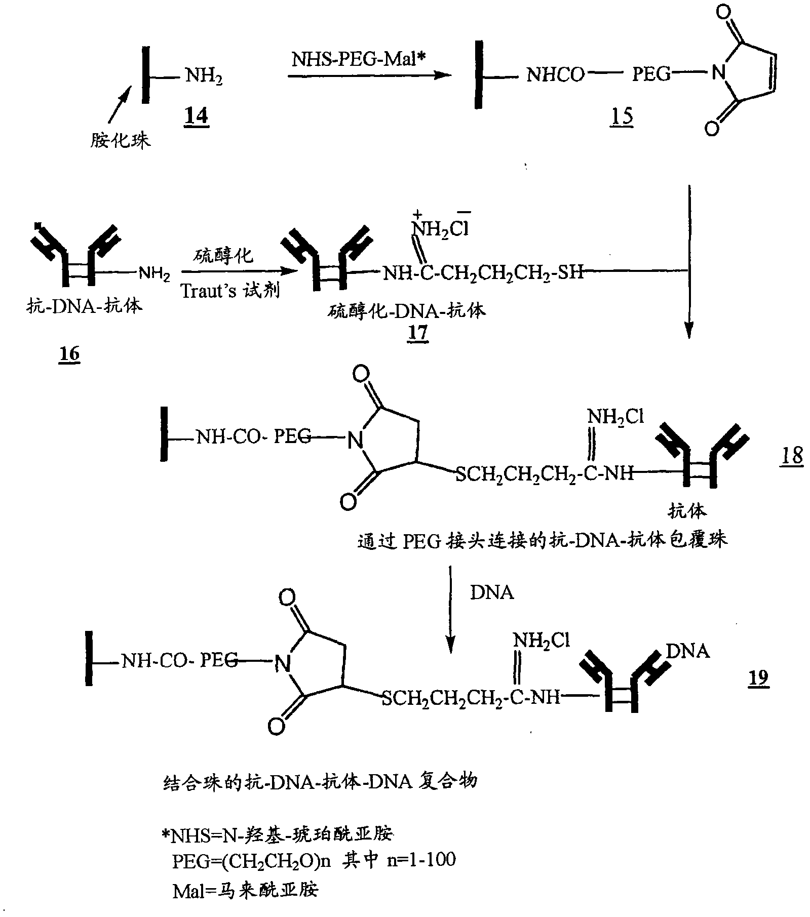
[0033] Example 1: Preparation of DNA binding beads

[0034] figure 1 , 2 and 3 outline the steps involved in the preparation of magnetic beads conjugated to anti-DNA-antibodies via IgG, protein-G and NHS-PEG-maleimide, respectively.

Embodiment 2

[0035] Example 2: Procedure for Isolating Fetal DNA from Maternal Blood

[0036] Beads coated with anti-DNA antibody 6 ( figure 1 ), 12( figure 2 ), or 18( image 3 ) to process 2ml of maternal blood. Use enough beads carrying at least 100 μg of anti-DNA antibody. Gently swirl the samples for 15 min at room temperature to ensure that the beads are well mixed with the blood. The samples were then placed in a magnetic separator for 1-2 minutes before removing the supernatant. The beads were then washed 3 times with 2M NaCl, 10 mM Tris.HCl, 1 mM EDTA (pH 7.0). The beads were then digested with proteinase K in 200 μl buffer (pH 8.0) containing 100 mM NaCl, 10 mM Tris.HCl, 25 mM EDTA, 1% SDS at 55°C for 1 hour. After proteinase K inactivation at 95 °C for 10 min, the supernatant was ethanol precipitated by adding 2 volumes of absolute ethanol and cooling the samples at −80 °C for 20 min. Wash the DNA pellet once with 90% ethanol.

Embodiment 3

[0037] Example 3: Determination of gender

[0038] The DNA from Example 2 was used as a template in PCR using primers and probes to determine fetal sex. After rinsing with 90% ethanol, the DNA pellet was dried, dissolved in 80 μl of water and analyzed for fetal sex by PCR. Using one or more TaqMan probes for Y chromosome sequence labeling, dual-labeled probes, 18-22 base oligonucleotides with a reporter fluorophore at the 5' end and a quencher fluorophore at the 3' end acid probe, and one or more primers to detect Y chromosome sequences.

[0039] SRY (Sex Determining Region Y) primers were used to target sex determining genes present on the Y chromosome of humans and other primates. The SRY gene encodes the testis determinant, also known as the SRY protein. Use FCY primers to target another commonly used marker in the Y chromosome. The β-hemoglobin gene, a housekeeping gene present in total DNA, was used as an internal control in each PCR reaction. As shown below, all 5...


PUM


Abstract

The present invention provides methods for isolating cell free nucleic acid, e.g., apoptotic or fetal nucleic acids and methods of detecting neoplastic cells or identifying the genetic composition of a fetus. The invention also provides magnetic particles comprising an anti-DNA antibody, and kits comprising the magnetic particles.

Description

Background technique [0001] Prenatal testing or screening is often done during pregnancy to determine the sex of the fetus or to detect genetic and / or chromosomal abnormalities in the fetus. To date, more than 4,000 genetic diseases have been recognized that are caused by one or more defective genes. Some examples include Cystic Fibrosis, Huntington's Disease, Beta Thalassaemia, Myotonic Dystrophy, Sickle Cell Anemia, Porphyria (Porphyria) and Fragile X Syndrome (Fragile-X-Syndrome). Chromosomal abnormalities are caused by aberrations in the number of chromosomes, duplication or deletion of chromosomal material, and by defects in chromosome structure. Examples of chromosomal abnormalities are trisomies such as trisomy 16, the leading cause of miscarriage in the first trimester, trisomy 21 (Down syndrome), trisomy 13 trisomy 13 (Patau syndrome), trisomy 18 (Edwards syndrome); Klinefelter's syndrome (47, XXY), (47, XYY) and (47, XXX); loss of chromosomes (monosomy), such as ...

Claims


Application Information

Patent Timeline
Patent Type & Authority Applications(China)
IPC IPC(8): C12Q1/68G01N33/53C07H21/00
CPCG01N33/54326C12Q1/6806C12Q1/6804G01N33/5308C12Q2525/204C12Q2522/101
Inventor 拉姆·巴特范文华
Owner NOVARTIS AG
Why Eureka
Try Eureka