Biomarker detection method and diagnostic kit for acute coronary syndrome

A technology of biomarkers and diagnostic kits, which is applied in the field of liquid-phase chip combined with parallel detection methods and diagnostic kits, can solve the problems of poor repeatability, insufficient sensitivity, and cumbersome operation of solid-phase biochip technology, and achieve high Sensitivity, stability, and high-throughput effects

Active Publication Date: 2011-05-18
MEDI GENETECH
View PDF2 Cites 20 Cited by
  • Summary
  • Abstract
  • Description
  • Claims
  • Application Information

AI Technical Summary

Problems solved by technology

However, these technologies can only detect one marker at a time, and the operation is cumbersome and the sensitivity is poor, which cannot really meet the needs of clinical diagnosis and detection.
However, solid-phase biochip technology has the disadvantages of poor reproducibility, insufficient sensitivity and cumbersome operation.

Method used


Image

  • Biomarker detection method and diagnostic kit for acute coronary syndrome
  • Biomarker detection method and diagnostic kit for acute coronary syndrome
  • Biomarker detection method and diagnostic kit for acute coronary syndrome

Examples


Embodiment 1

[0044] Example 1: Liquid-phase chip combined parallel detection method for three markers of acute coronary syndrome

[0045] The specific detection method includes the following steps:

[0046] 1. Activation of the required microspheres:

[0047] 1.1 Vortex the microsphere storage solution at full speed for at least 3 minutes to form a uniform microsphere suspension;

[0048] 1.2 Weigh 10 mg of EDC and S-NHS into two centrifuge tubes;

[0049] 1.3 Dissolve in deionized water to make the final concentration 50mg / ml;

[0050] 1.4 Take 1ml of the microsphere suspension and centrifuge at 10000g for 3min, carefully remove the supernatant;

[0051] 1.5 Add 80 μl of activation buffer to resuspend the microspheres;

[0052] 1.6 Add 10 μl of EDC solution (50 mg / ml) and 10 μl of S-NHS solution (50 mg / ml) respectively, mix well, and incubate at room temperature (15-25° C.) in the dark and shake for 20 minutes.

[0053] 2. Coupling of corresponding capture antibodies to activated mic...

Embodiment 2

[0099] Example 2: Liquid chip combined parallel detection method for six acute coronary syndrome markers The specific detection method, steps 1-3 are the same as in Example 1.

[0100] 4. Configuration of Antigen Standards

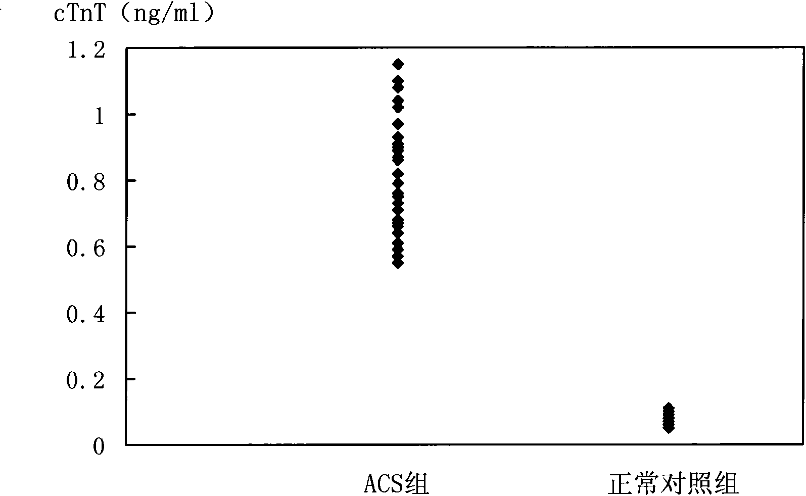
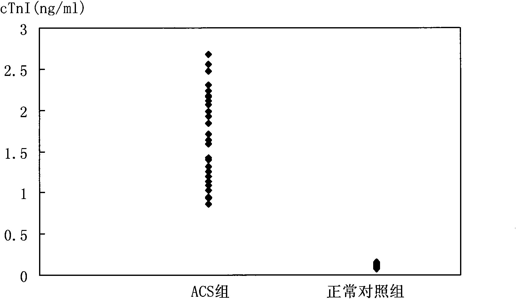
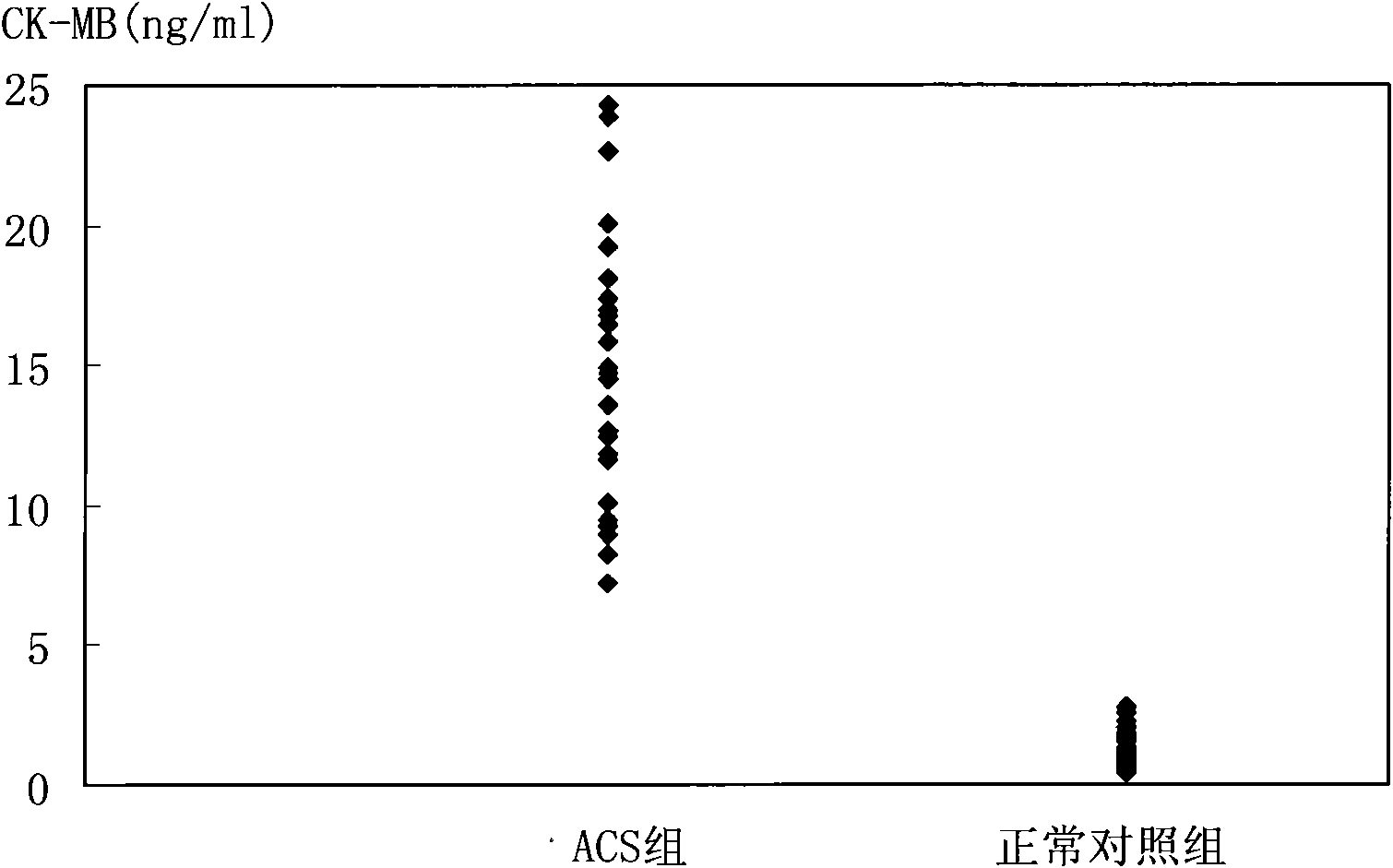
[0101] cTnT and cTnI are prepared according to the concentration of 31.25, 6.25, 1.25, 0.25, 0.05, 0.01, 0ng / ml, CK-MB and H-FABP are prepared according to the concentration of 312.5, 62.5, 12.5, 2.5, 0.5, 0.1, 0ng / ml For preparation, Myo and MPO were prepared at concentrations of 3125, 625, 125, 25, 5, 1, and 0 ng / ml, and the marker mixtures were labeled as STD6, ​​STD5, STD4, STD3, STD2, STD1, and STD0, respectively.

[0102] 5. Preparation of microsphere mixture (mixture I) coupled with capture antibody

[0103] Take the microspheres coated with the capture antibodies of the 6 ACS markers, as follows: cTnT capture antibody microspheres 11, cTnI capture antibody microspheres 15, CK-MB capture antibody microspheres 21, H-FABP capture antibody microsphere...


PUM

PropertyMeasurementUnit
diameteraaaaaaaaaa

Abstract

The invention discloses a biomarker detection method and a diagnostic kit for acute coronary syndrome. The detection method is characterized in that: a plurality of biomarkers in the same sample can be detected once, namely, a biotin-marked detection antibody-acute coronary syndrome biomarker-capture antibody-bead tetragenous complex is formed by the preparation of a liquid chip, and is combined with streptavidin-phycoerythrin (PE) to detect fluorescence signals of different beads, thereby determining the existence of different acute coronary syndrome biomarkers in the sample to be detected and the acute coronary syndrome biomarker content of the sample. The invention also discloses components of the diagnostic kit. The method and the kit provided by the invention have the advantages of high sensitivity, high flux, high detection speed, detection accuracy and the like, and can simultaneously realize the qualitative and quantitative detection of the plurality of acute coronary syndromebiomarkers.

Description

technical field [0001] The invention relates to an in vitro diagnostic detection method and a diagnostic kit, in particular to a liquid-phase chip combined parallel detection method and a diagnostic kit for biomarkers of various acute coronary syndromes. Background technique [0002] Acute Coronary Syndrome (ACS) is a group of clinical syndromes characterized by instability of coronary atherosclerotic plaques, acute myocardial ischemia as a common feature, and chest pain as a typical symptom. ACS is the main reason for most patients with cardiovascular disease to see a doctor and die suddenly, which seriously endangers human life and health. ACS biomarkers (hereinafter referred to as "ACS markers") can not only accurately diagnose acute coronary syndrome and evaluate its severity, but also guide clinical treatment and efficacy monitoring, and judge the prognosis of acute coronary syndrome. Therefore, the combined parallel detection of various types of ACS markers has become...

Claims


Application Information

Patent Timeline
Patent Type & Authority Applications(China)
IPC IPC(8): G01N21/76G01N21/64G01N33/68G01N33/70
Inventor 邵棠孙黎吴杰
Owner MEDI GENETECH
Why Eureka
Try Eureka