
Optical three-dimensional imaging method based on biological tissue specificity

A biological tissue, three-dimensional imaging technology, applied in the field of optics, can solve the problems of inability to achieve accurate, fast, high-resolution reconstruction of targeted targets, without considering the sparse characteristics of the biological area of ​​measurement data, etc., to achieve rapid reconstruction, Overcome the inability of imaging accuracy and imaging efficiency to balance, and overcome the effect of insufficient imaging resolution

Active Publication Date: 2012-03-28
GUANGZHOU BLT INSTR & METER
View PDF5 Cites 20 Cited by
  • Summary
  • Abstract
  • Description
  • Claims
  • Application Information

AI Technical Summary

Problems solved by technology

However, this method only uses a single optimization strategy to solve the objective function, and cannot achieve the optimal balance between imaging accuracy and imaging speed under different problem scales.
In addition, the method does not take into account the sparse nature of the measurement data relative to the area of ​​the organism
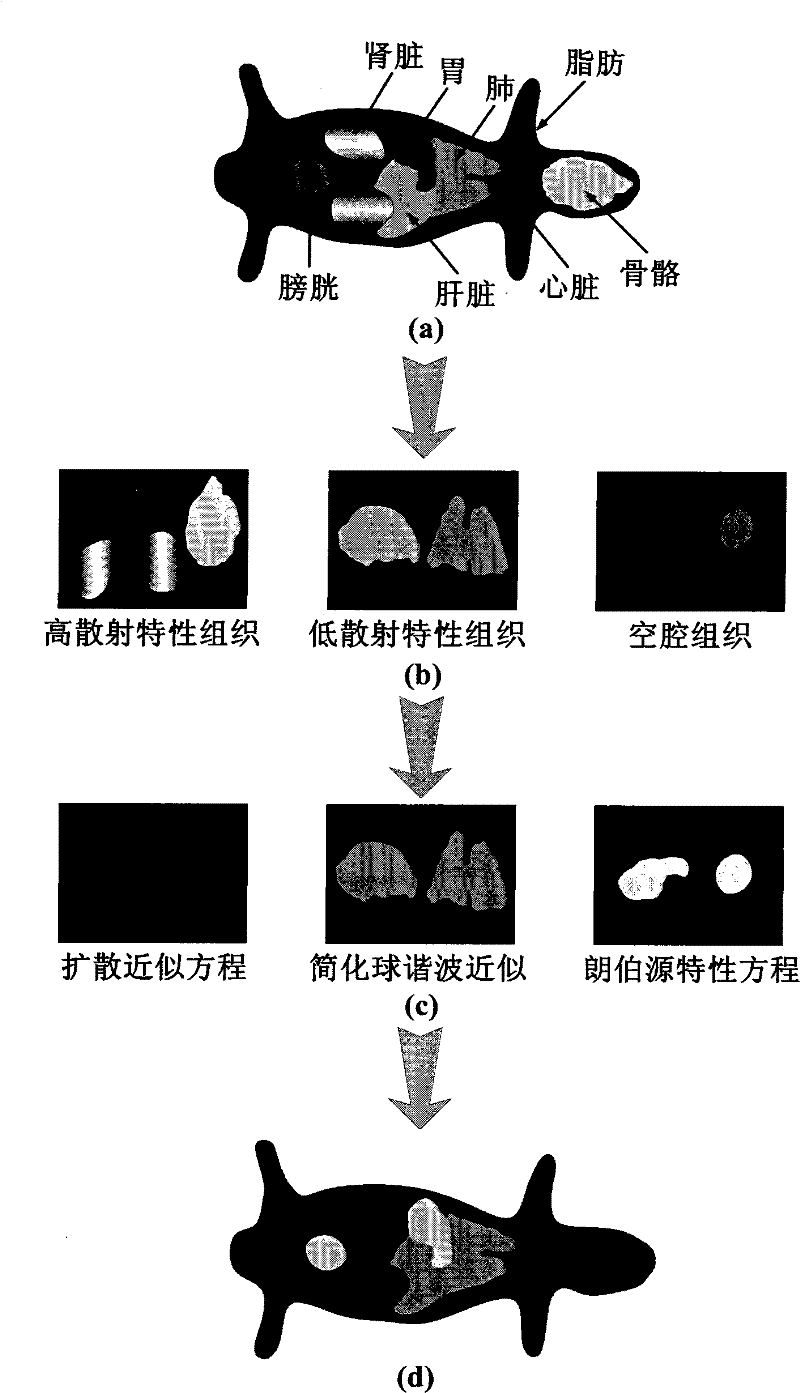
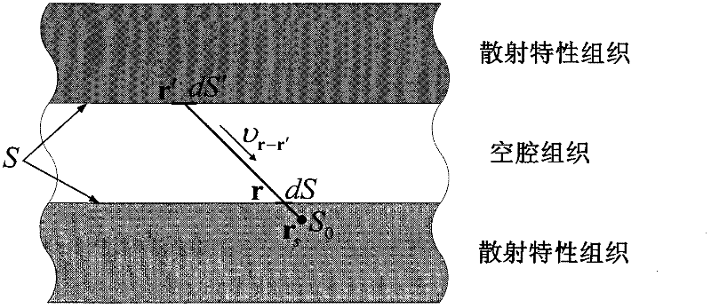
[0007] In summary, for complex organisms with irregular anatomical structures and tissues with various scattering characteristics, the existing optical 3D imaging methods based on a single approximation equation or mixed light transport equation and optical 3D imaging based on a single optimization strategy None of these methods can achieve accurate, rapid and high-resolution reconstruction of the target in vivo

Method used


Image

  • Optical three-dimensional imaging method based on biological tissue specificity
  • Optical three-dimensional imaging method based on biological tissue specificity
  • Optical three-dimensional imaging method based on biological tissue specificity

Examples


Embodiment Construction

[0057] The present invention will be further described below in conjunction with the accompanying drawings.

[0058] refer to figure 1 , the concrete steps of the present invention are as follows:

[0059] Step 1, collect data

[0060] Using the multi-modal molecular imaging system, multi-angle fluorescence data for optical three-dimensional imaging, multi-angle laser data for reconstruction of optical characteristic parameters, and computed tomography projection data for obtaining animal body anatomy are sequentially collected.

[0061] For the collection of multi-angle fluorescence data, control the imaging body to rotate at a certain angle at equal intervals, generally no more than 90° (90° is selected in this example), and use the CCD camera in the multi-modal molecular imaging system to collect fluorescence data from no less than four angles (four angles in this example). Continue to rotate the imaging volume back to the position where the fluorescence data was origina...


PUM


Abstract

The invention discloses an optical three-dimensional imaging method based on biological tissue specificity. The problem of the prior art that the accurate and quick optical three-dimensional imaging cannot be performed on a complex organism having an irregular anatomical structure and various scattering characteristic tissues is solved. According to the method, a target function is established onthe basis of an optical transmission mixed mathematical model for the biological tissue specificity and a completely thinning regularizing method, and a mixed optimizing method based on a task orientation is used for solving, thereby realizing the optical three-dimensional imaging for an internal targeted object. The method comprises the following steps: collecting a data; preprocessing the data;acquiring the anatomical structure; acquiring a surface optical data; reestablishing optical characteristic parameters; establishing an optical transmission model; establishing a system equation; establishing the target function; solving the target function; and displaying a result. The optical three-dimensional imaging method has the advantage that the accurate, quick and high-resolution reconstruction for a light source in the complex organism can be realized, and the method can be applied to the field of internal optical three-dimensional imaging.

Description

technical field [0001] The invention belongs to the field of optical technology, and further relates to an optical three-dimensional imaging method based on biological tissue specificity in the field of optical molecular imaging technology. This method can be applied to small animal experiments and pre-clinical experiments such as gene expression, tumor detection, drug development and efficacy evaluation to obtain the position and intensity information of the target. Background technique [0002] As an emerging optical imaging technology, optical three-dimensional imaging is to reconstruct living organisms based on accurate light transmission models in biological tissues by fusing multi-angle optical signals measured on the surface of organisms, anatomical structures of organisms, and tissue optical parameter information. The position and intensity distribution information of the target in the body, among which, the accurate description of the light transmission process in t...

Claims


Application Information

Patent Timeline
IPC IPC(8): G06T17/00G06T19/00
Inventor 梁继民陈雪利田捷屈晓超陈多芳朱守平赵恒
Owner GUANGZHOU BLT INSTR & METER
Learn More