
Method for simply preparing DiI (1,1'-dioctadecyl-3,3,3',3'-tetramethylindocarbocyanine perchlorate) microscopic particles and marking neurons

A production method and neuron technology, applied in the field of biomedical experiments, can solve the problems of high technical difficulty, complex production process and high cost, and achieve the effects of simple operation, simple production and low cost

Inactive Publication Date: 2012-04-11
FOURTH MILITARY MEDICAL UNIVERSITY
View PDF6 Cites 1 Cited by
  • Summary
  • Abstract
  • Description
  • Claims
  • Application Information

AI Technical Summary

Problems solved by technology

The disadvantage of this technology is that the production process of "bullets" is complicated, the cost is high, the technology is difficult, and the "gene gun" is required

Method used


Image

  • Method for simply preparing DiI (1,1'-dioctadecyl-3,3,3',3'-tetramethylindocarbocyanine perchlorate) microscopic particles and marking neurons
  • Method for simply preparing DiI (1,1'-dioctadecyl-3,3,3',3'-tetramethylindocarbocyanine perchlorate) microscopic particles and marking neurons
  • Method for simply preparing DiI (1,1'-dioctadecyl-3,3,3',3'-tetramethylindocarbocyanine perchlorate) microscopic particles and marking neurons

Examples


Embodiment Construction

[0031] The present invention will be described in further detail below in conjunction with the accompanying drawings and specific embodiments given by the inventor.

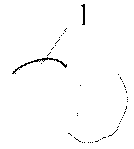
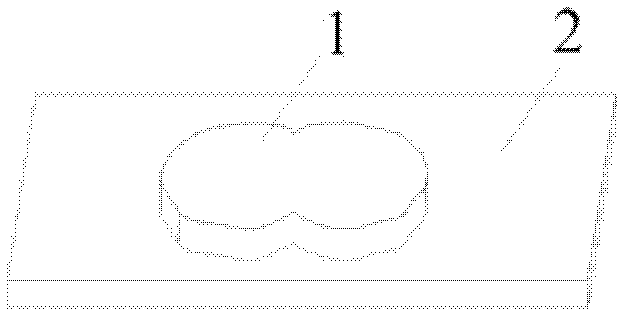
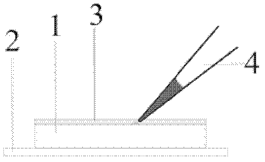
[0032] see Figure 1 to Figure 4 As shown, the method for labeling neurons with DiI microparticles includes 4 steps, namely, preparation of brain slices, preparation of DiI particles, embedding of DiI particles, and incubation of brain slices.

[0033] (1) Brain slice production.

[0034] The brain slices used to label neurons can be live or fixed brain slices, which are performed according to the brain slice patch clamp or immunohistochemical brain slice preparation techniques. Both brain slices must be sliced ​​with a vibrating microtome with a thickness of 200-350 microns. Live brain slices were excised and incubated in oxygenated artificial cerebrospinal fluid (aCSF), while fixed isolated brain slices were placed in paraformaldehyde fixative solution with a mass volume concentration of 0.015-0.02 g / ml.

[...


PUM


Abstract

The invention relates to a method for simply preparing DiI (1,1'-dioctadecyl-3,3,3',3'-tetramethylindocarbocyanine perchlorate) microscopic particles and marking neurons, wherein the method for simply preparing DiI microscopic particles comprises the following steps of: dissolving DiI particles in absolute ethanol or dimethyl sulfoxide to form a DiI solution of which the mass volume concentration is 0.01-0.1g / ml; adding the DiI solution of which the mass volume concentration is 0.01-0.1g / ml into artificial cerebrospinal fluid or phosphate buffer of which the concentration is 0.1mol / L; and enabling the DiI solution to quickly float on the artificial cerebrospinal fluid or phosphate buffer in a film form, and moving and diffusing all around to precipitate DiI microscopic particles of which the particle sizes are 3-10 micrometers. The method provided by the invention is simple in operation and low in cost.

Description

【Technical field】 [0001] The invention belongs to the field of biomedical experiments, and is suitable for marking neurons in specific brain regions on isolated brain slices of animals under physiological and pathological conditions, so as to microscopically observe and qualitatively observe the neuron morphology and its changes, the number of neuron dendritic spines, etc. and quantitative analysis. 【Background technique】 [0002] Use fluorescent dyes to mark neurons in specific brain parts, and use fluorescence microscopy or laser confocal microscopy to conduct morphological qualification or quantitative research on dendritic spines on these labeled neurons. The pathology of neurological diseases is an indispensable biomedical experimental technique, which is required to be applied in almost all laboratories conducting neuroscience research. Lipophilic carbocyanine dyes, which can clearly mark neuronal morphological details, are very important and commonly used fluorescent...

Claims


Application Information

Patent Timeline
IPC IPC(8): C09B67/48G01N21/84
Inventor 张富兴李云庆
Owner FOURTH MILITARY MEDICAL UNIVERSITY
Learn More