Tumor cell migration dynamics monitoring method based on microfluidic chip
A microfluidic chip, tumor cell technology, applied in biochemical equipment and methods, microbial determination/inspection, enzymology/microbiology devices, etc., can solve problems such as being in the blank stage
- Summary
- Abstract
- Description
- Claims
- Application Information
AI Technical Summary
Problems solved by technology
Method used
Image
Examples
Embodiment 1
[0034] Cell Migration of HepG-2 Cells in Different Concentrations of Collagen
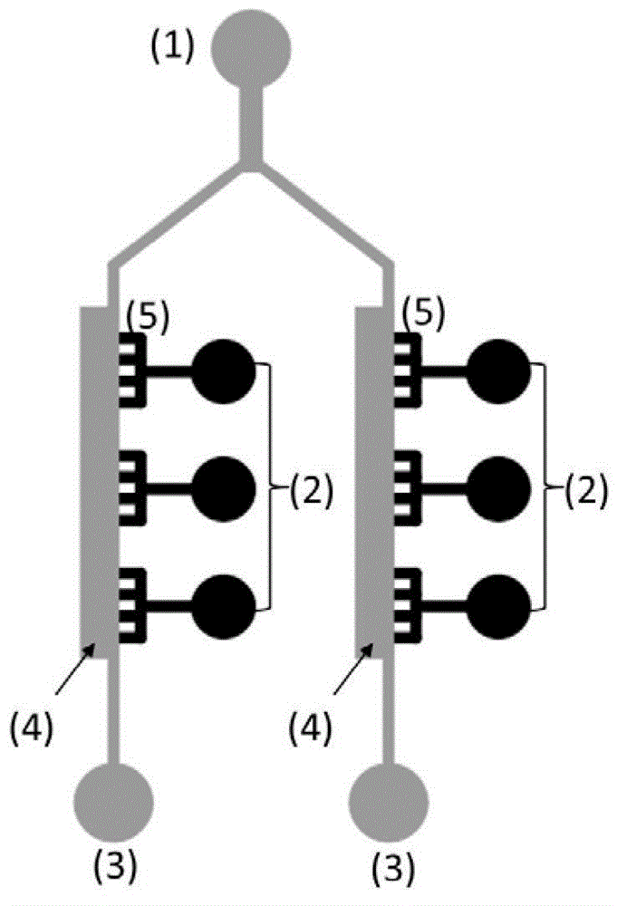
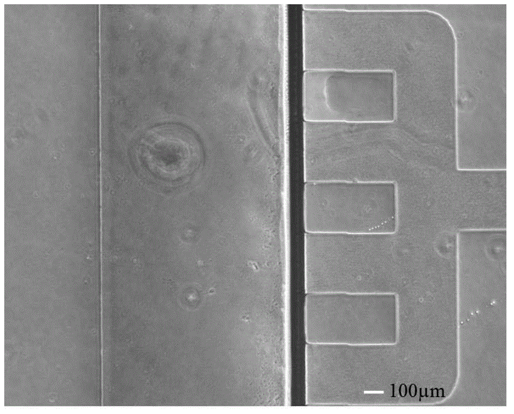
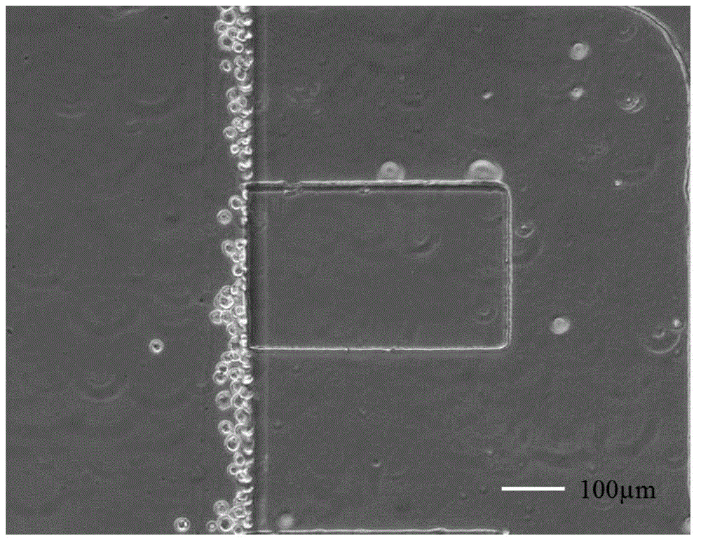
[0035] Three different concentrations of collagen working solutions were prepared, the concentrations were 1mg / mL, 2.5mg / mL, and 5mg / mL, and the distribution of collagen filaments with different concentrations was as follows: Figure 4 shown. Using the microfluidic chip designed and manufactured by the laboratory, the configuration is as follows: figure 1 shown. After perfusing the collagen into the chip and incubating for 30 minutes, a clear interface between the collagen and the two-dimensional plane can be seen ( figure 2 ). Add 10 μL of 1 x 10 from the cell inlet pool 6 / mL HepG-2 cell suspension, set the chip upright for 10 minutes, so that the cells can attach to the side of the collagen, such as image 3 shown. Take pictures to record the initial position of the cells, change the medium every 24 hours, and take pictures to record the cell movement. Counting the area of HepG-2 cells...
Embodiment 2
[0037] Real-time monitoring of migration of SMMC-7721 in 2.5mg / mL collagen
[0038] Using the microfluidic chip designed and manufactured by the laboratory, the configuration is as follows: figure 1shown. After the 2.5mg / mL collagen was poured into the chip for coagulation, the same cell inoculation and culture methods as in Example 1 were adopted. 24 hours after the cells were seeded on the chip, the culture dish with the chip fixed was put into the stage incubator. Adjust the focal length and shooting parameters, take a picture every 60 minutes, and record the cell movement position and shape changes in real time by taking pictures under the microscope. The shooting time is 19 hours. The shooting results are as follows: Figure 8 shown.
Embodiment 3
[0040] After adding relaxin D, SMMC-7721 migration movement analysis in 2.5mg / mL collagen.
[0041] Using the microfluidic chip designed and manufactured by the laboratory, the configuration is as follows: figure 1 shown. After the 2.5mg / mL collagen was poured into the chip for coagulation, the same cell inoculation and culture methods as in Example 1 were adopted. The relationship between the area of SMMC-7721 cells migrating into collagen and the culture time is shown in Figure 9 shown. After the cells were seeded on the chip for 24 hours, 1 μg / mL relaxin D was added to the culture medium to stimulate the cells, and the morphological changes and migration behavior of the cells under the action of relaxin D for 24 hours were recorded. The results are as follows: Figure 10 As shown, the relationship between the migration area of SMMC-7721 cells and the culture time is shown in Figure 11 shown.
PUM
 Abstract
Description
Claims
Application Information
 - R&D
- Intellectual Property
- Life Sciences
- Materials
- Tech Scout
- Unparalleled Data Quality
- Higher Quality Content
- 60% Fewer Hallucinations
Browse by: Latest US Patents, China's latest patents, Technical Efficacy Thesaurus, Application Domain, Technology Topic, Popular Technical Reports.
© 2025 PatSnap. All rights reserved.Legal|Privacy policy|Modern Slavery Act Transparency Statement|Sitemap|About US| Contact US: help@patsnap.com


