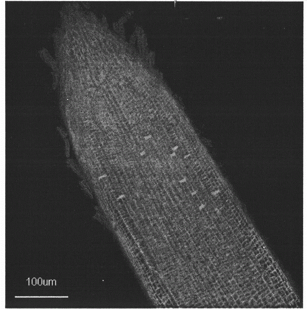
Method for in vitro detection of transport of Magnaporthe grisea effector proteins in root tissues of rice
A technology of blast fungus and effector protein, which is applied in vitro to detect the translocation of blast fungus effector protein in rice root tissue, and achieves the effect of simple method
- Summary
- Abstract
- Description
- Claims
- Application Information
AI Technical Summary
Problems solved by technology
Method used
Image
Examples
Embodiment 1
[0015] 5.0mg / ml of GFP fusion protein was treated at 0.5cm of rice root tip for 12h, rinsed with sterilized water for 4h, and the transport of GFP fusion protein in rice root tissue was directly observed by confocal microscope.
[0016] The specific method is:
[0017] The expression product of the rice blast fungus effector protein gene (the process of obtaining the expression product by using the method of IPTG chemical induction will not be repeated) was centrifuged at 4°C to collect the bacterial cells, and the freshly prepared buffer solution (20mM Tris-HCl, pH7.4 ; 200mM NaCl; 1mM EDTA; 10mM β-mercaptoethanol) suspended cells. The supernatant was collected by centrifugation after ultrasonography to disrupt the cells in an ice-water bath. The prokaryotic expression product was purified using a GST purification column (purchased from Amersia Company), and the purified expression product was treated with the root of the susceptible rice variety Lijiang Xintuan Heigu at a c...
Embodiment 2
[0021] 5.0mg / ml of GFP fusion protein was treated at 0.5cm of rice root tip for 24h, rinsed with sterilized water for 4h, and the transport of GFP fusion protein in rice root tissue was directly observed by confocal microscope.
[0022] Centrifuge the expression product of the effector protein gene of Magnaporthe grisea to collect the cells, and suspend the cells with freshly prepared buffer (20mM Tris-HCl, pH7.4; 200mM NaCl; 1mM EDTA; 10mM β-mercaptoethanol) . The supernatant was collected by centrifugation after ultrasonography to disrupt the cells in an ice-water bath. The treatment is to treat the root tissue of the susceptible rice variety Lijiang Xintuan Heigu at a concentration of 5.0mg / ml (including 25mM MES) after purification of the prokaryotic expression product of the gene for 24 hours, rinse with sterilized water for 4 hours, and observe the effector protein with a confocal microscope Gene transport in rice root tissue. As a control, after the prokaryotic expres...
Embodiment 3
[0026] The GFP fusion protein of 5.0 mg / ml was treated at the root tip of rice at 0.5 cm for 24 hours, rinsed with sterilized water for 4 hours, and the translocation of GFP fusion protein in rice root tissue was directly observed by confocal microscopy.
[0027] Centrifuge the expression product of the effector protein gene of Magnaporthe grisea to collect the cells, and suspend the cells with freshly prepared buffer (20mM Tris-HCl, pH7.4; 200mM NaCl; 1mM EDTA; 10mM β-mercaptoethanol) . The supernatant was collected by centrifugation after ultrasonography to disrupt the cells in an ice-water bath. The treatment is to treat the root tissue of the susceptible rice variety Lijiang Xintuan Heigu at a concentration of 5.0mg / ml (including 25mM MES) after purification of the prokaryotic expression product of the gene for 36 hours, rinse with sterilized water for 4 hours, and observe the effector protein with a confocal microscope Gene transport in rice root tissue. As a control, a...
PUM
 Abstract
Description
Claims
Application Information
 - R&D
- Intellectual Property
- Life Sciences
- Materials
- Tech Scout
- Unparalleled Data Quality
- Higher Quality Content
- 60% Fewer Hallucinations
Browse by: Latest US Patents, China's latest patents, Technical Efficacy Thesaurus, Application Domain, Technology Topic, Popular Technical Reports.
© 2025 PatSnap. All rights reserved.Legal|Privacy policy|Modern Slavery Act Transparency Statement|Sitemap|About US| Contact US: help@patsnap.com


