
X-ray image diagnosis device, and medical image processing program and method

An image diagnosis and X-ray technology, applied in image data processing, image data processing, instruments used for radiological diagnosis, etc., can solve problems such as pixel pitch and grid density interference

Inactive Publication Date: 2012-11-07
HITACHI LTD
View PDF7 Cites 13 Cited by
  • Summary
  • Abstract
  • Description
  • Claims
  • Application Information

AI Technical Summary

Problems solved by technology

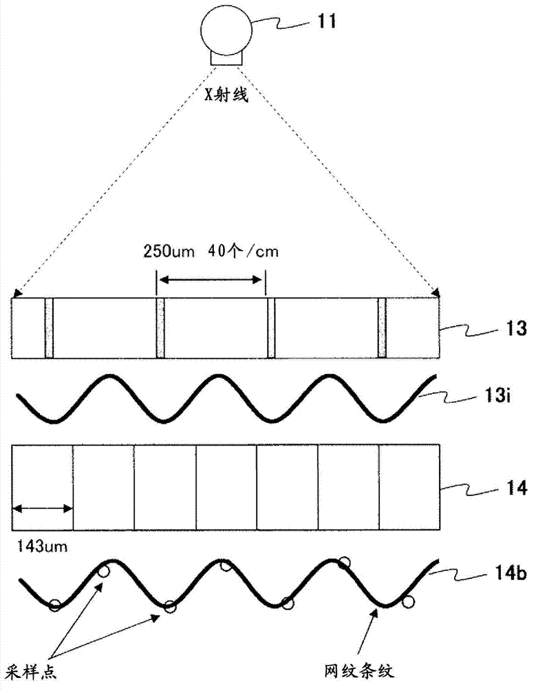
However, the image generated by the X-ray plane detector samples the image signal two-dimensionally, so the pixel pitch and the grid density interfere, causing aliasing (moire)

Method used


Image

  • X-ray image diagnosis device, and medical image processing program and method
  • X-ray image diagnosis device, and medical image processing program and method
  • X-ray image diagnosis device, and medical image processing program and method

Examples


Embodiment Construction

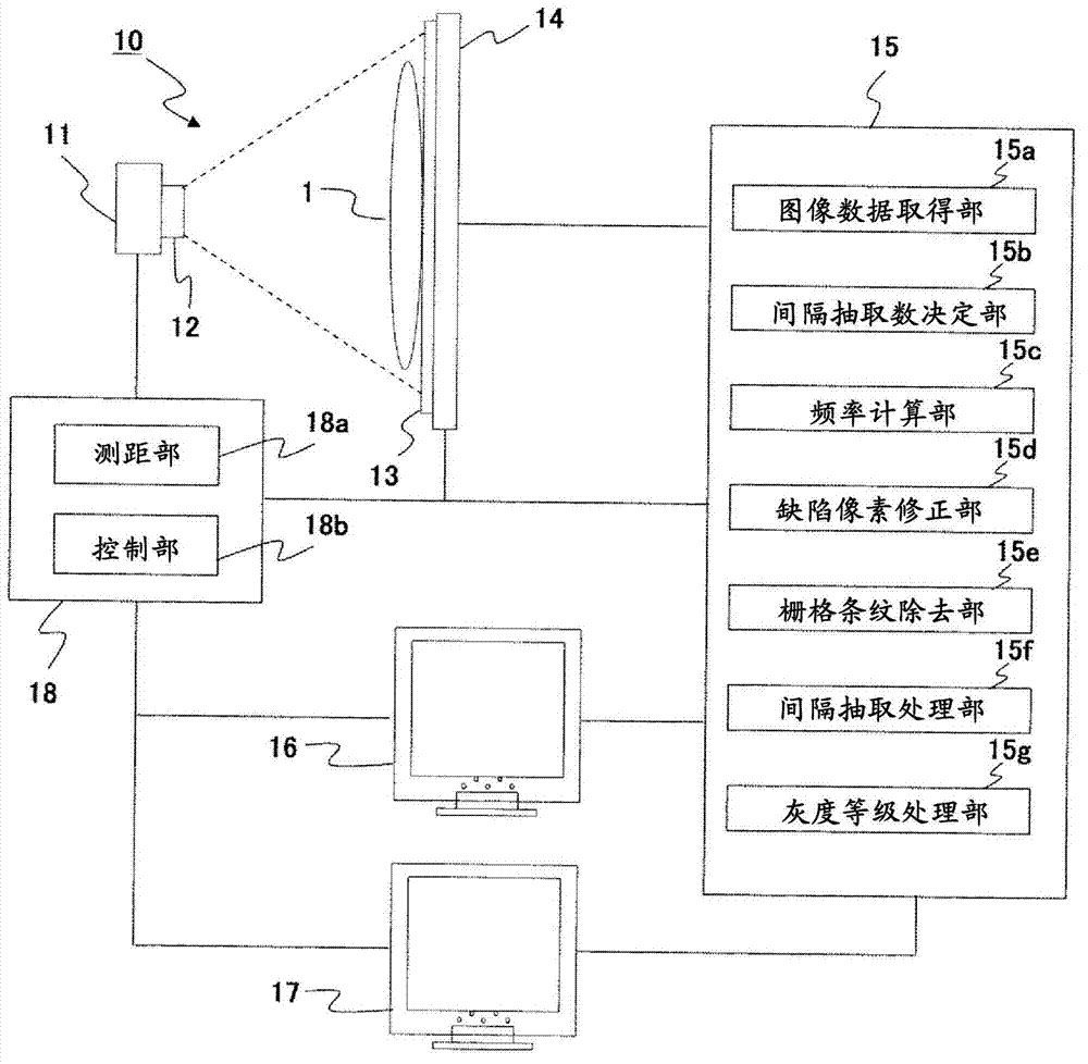
[0025] Hereinafter, embodiments of the present invention will be described with reference to the drawings. figure 1 It is a schematic diagram showing a schematic configuration of the X-ray image diagnostic apparatus 10 according to the present embodiment.

[0026] The X-ray image diagnostic apparatus 10 includes: an X-ray tube 11 for irradiating X-rays to the subject 1; an X-ray diaphragm 12 for limiting the irradiation of the X-rays irradiated from the X-ray tube 11 to the subject 11; The grid 13 of the scattered X-ray generated by 1; the X-ray plane detector 14 that is arranged opposite to the X-ray tube 11 and detects the X-ray transmission of the subject 1; the image data output from the X-ray plane detector 14 is implemented Image processing device 15 for image processing such as defect pixel correction, grid stripe removal processing, grayscale processing, etc.; preview display device 16 for displaying preview (photography confirmation) image data generated by image proc...


PUM


Abstract

In order to remove grid stripes from image data subjected to thinning processing without causing artifacts, image data obtained by capturing an image of a subject by an X-ray image diagnosis device provided with a grid is acquired (S11), the thinning number is determined (S13), the frequency of grid stripes included in the image data is calculated on the basis of the determined thinning number (S21), defective pixel correction corresponding to the calculated frequency of the grid stripes is performed (S41-S45), and the grid stripes are removed from the corrected image data (S51-S55).

Description

technical field [0001] The present invention relates to an X-ray image diagnostic device, a medical image processing program, and a method, and particularly to improving the image quality of an image obtained by X-ray imaging using a grid for removing scattered X-rays. Background technique [0002] Conventionally, a grid is arranged on an X-ray plane detector to prevent imaging of scattered X-rays generated inside an object. However, since the image generated by the X-ray flat surface detector samples the image signal two-dimensionally, the pixel pitch and the grid density interfere, and aliasing (moire) occurs. As a technique for reducing this moire, for example, Patent Document 1 discloses an image processing method in which a frequency band of a moire component is calculated from the pixel size of an X-ray plane detector and the grid density of a grid, Select the grid where the texture appears in the high frequency band, perform filter processing or Fourier transform, re...

Claims


Application Information

Patent Timeline
IPC IPC(8): A61B6/00G06T1/00G06T5/00
CPCG06T2207/10116G06T2207/30061G06T5/002A61B6/4291A61B6/4452A61B6/5258G06T5/70
Inventor 竹之内忍铃木克己
Owner HITACHI LTD
Learn More